Download
1 / 34
350 likes | 1.1k Views
A. Nervous system divisions B. Functional anatomy of nervous tissue 1. Neuroglia a. Types of neuroglia b. Myelination 2. Neurons a. Parts of a neuron b. Classification of

E N D
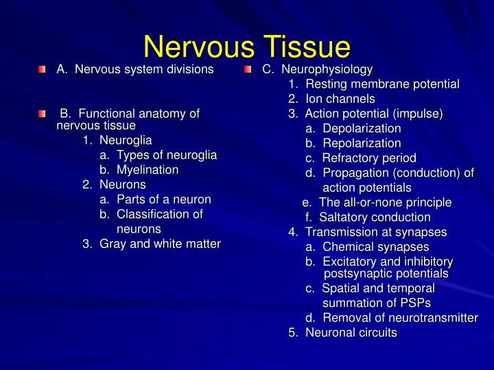
A. Nervous system divisions B. Functional anatomy of nervous tissue 1. Neuroglia a. Types of neuroglia b. Myelination 2. Neurons a. Parts of a neuron b. Classification of neurons 3. Gray and white matter C. Neurophysiology 1. Resting membrane potential 2. Ion channels 3. Action potential (impulse) a. Depolarization b. Repolarization c. Refractory period d. Propagation (conduction) of action potentials e. The all-or-none principle f. Saltatory conduction 4. Transmission at synapses a. Chemical synapses b. Excitatory and inhibitory postsynaptic potentials c. Spatial and temporal summation of PSPs d. Removal of neurotransmitter 5. Neuronal circuits Nervous Tissue
The nervous system is the body's control center and communicates network. It serves three broad functions: • 1. senses changes in the environment • 2. integrates and interprets • 3. responds
1. central nervous system a. brain b. spinal cord 2. peripheral nervous system a. somatic division b. autonomic division (1) sympathetic vs (2) parasympathetic Nervous Systems Divisions
Neuroglia • 1. astrocytes • 2. oligodendrocytes • 3. microglia • 4. ependyma • 5. neurolemmocytes (Schwann cells) • 6. satellite cells
Neuron • 1. cell body a. nucleus b. nucleolus c. Nissl substance • 2. dendrite • 3. axon a. axon hillock b. trigger zone c. axon collaterals d. telodendrion e. end bulbs
Myelination • 1. PNS = neurolemmocytes CNS = oligodendrocytes • 2. process • 3. myelin sheath • 4. neurolemma • 5. nodes of Ranvier
Structural Classification of Neurons • 1. multipolar neuron • 2. bipolar neuron • 3. unipolar neuron a. central process b. receptor
Functional Classification of Neurons • 1. sensory (afferent) neurons • 2. association neurons (interneurons) • 3. motor (efferent) neurons
Some Terminology • 1. nerve fiber • 2. nerve vs. tract • 3. tract • 4. ganglion vs. nucleus (center) • 5. gray matter • 6. white matter Gray matter White matter
Neurophysiology Communication by neurons depends upon two basic properties of their cell membranes: • 1. There is an electrical voltage, called the resting membrane potential, across the cell membrane. • 2. Their cell membranes contain a variety of ion channels (pores) that may be open or closed.
Resting Membrane Potential • 1. build-up of ions • 2. separation of charges = potential energy (mV) • 3. membrane potential = -70 mV • 4. polarized membrane +++++ +++++ -------- ---------
Two Main Factors Contribute to the RMP • 1. distribution of ions across the cell membrane. a. extracellular fluid is rich in Na+ and Cl- b. intracellular fluid is rich in K+ and anions such as organophosphates and proteins • 2. relative permeability of the cell membrane to Na+ and K+ a. moderately permeable to K+ and Cl- b. slightly permeable to Na+ c. impermeable to the intracellular anions
Ion Channels(Pores) • 1. non-gated (leakage) • 2. gated (regulated) a. open in response to stimulus (chemical, voltage change, light, mechanical) b. excitable cells have them c. found in trigger zones
How do chemically gated ion channels open? Na+ Na+ Na+ Na+ Na+ ACh Na+ Na+ extracellular fluid extracellular fluid Na+ Na+ Na+ Na+ ACh Na+ +++++++ +++++++++++++ +++++++ +++++++++++++ Na+ cell membrane cellmembrane Na+ Na+ Na+ - - - - - - - - - - - - - - - - - - -- - - -- - - - - - - - - - - - - - - -- Na+ Na+ CHEMICALLY-GATED Na+ CHANNEL closed CHEMICALLY-GATED Na+ CHANNEL open intracellular fluid intracellular fluid
How do voltage-gated channels open? Voltage Na+ Na+ Na+ Na+ Na+ Na+ Na+ extracellular fluid extracellular fluid Na+ Na+ Na+ Na+ Na+ +++++++ +++++++++++++ +++++++ +++++++++++++ Na+ cell membrane cell membrane Na+ Na+ Na+ - - - - - - - - - - - - - - - - - - -- - - - - - - - - - - - - - - - - - - -- Na+ Na+ 3. Mechanically-gated channels, light-gated channels VOLTAGE-GATED Na+ CHANNEL closed VOLTAGE GATED Na+ CHANNEL open intracellular fluid intracellular fluid
Action Potential-Depolarization • 1. threshold stimulus • 2. voltage-gated Na+ channels open • 3. Na+ influx (-70 ---> +30 mV) _____________________________ • 4. positive feedback
Action Potential-Repolarization • 1. voltage-gated K+ channels open • 2. K+ efflux (-90 <--- +30 mV) • 3. hyperpolarization • 4. Na-K pumps restore ions
Refractory Period • 1. absolute • 2. relative
POSITIVE FEEDBACK OF AN ACTION POTENTIAL CONTROLLED CONDITION A stimulus or stress disrupts membrane homeostasis by causing a threshold depolarization NO RETURN TO HOMEOSTASIS Opening of the voltage-gated Na+ channels causes depolarization in adjacent membrane, opening more voltage-gated Na+ channels RECEPTOR The receptors in this case are voltage-gated Na+ channels in their resting state EFFECTORS Voltage-gated Na+ channels are also effectors. Threshold depolarization causes shape changes in the channel CONTROL CENTER The shape of the voltage-gated Na+ channel depends on membrane voltage change
All of None Principle Each time an action potential is formed, it has a constant and maximum strength for the existing conditions.
Continuous Conduction • 1. trigger zone to synapse • 2. propagation • 3. one direction only
Saltatory Conduction • 1. myelin sheath • 2. nodes of Ranvier • 3. "jumping" impulse • 4. 0.5 vs 130 m/sec • 5. energy conservation
Transmission at Synapses • 1. "synapsis" means connection • 2. synapses integrate and filter information • 3. signals are transmitted or inhibited • 4. presynaptic vs postsynaptic neurons
Chemical Synapses • 1. arrival of action potential • 2. Ca++ influx • 3. synaptic vesicle rupture • 4. NT release • 5. NT diffusion • 6. NT/receptor interaction • 7. postsynaptic potential
Postsynaptic Potentials can be excitatory or inhibitory • 1. excitatory (EPSP) a. facilitation b. summation (1) spatial (2) temporal • 2. inhibitory (IPSP) a. hyperpolarization
Facilitation and Summation • 1. spatial • 2. temporal ___________ • net effects • 1. facilitation • 2. summation (impulses) • 3. inhibition (hyperpolarization)
EXAMPLE OF SYNAPTIC INTEGRATION postsynaptic neuron inhibitory presynaptic neuron excitatory presynaptic neuron Threshold= +3
TYPES OF NEUROTRANSMITTERS Acetylcholine Most common neurotransmitter; In a class by itself chemically; Mostly excitatory, depending on location and function; Brain, spinal cord, neuromuscular and neuroglandular synapses of the periphery Excitatory amino acids Glutamate – 75% of excitatory synapses in brain Asparate – spinal cord Inhibitory amino acids Glycine – most common in spinal cord GABA (gamma amino butyric acid) – most common in brain Monoamines (biogenic amines) Catecholamines – norepinephrine, epinephrine, dopamine Other amines – serotonin, histamine Neuropeptides – substance P, enkephalines and endorphins, cholecystokinin
Removal of Neurotransmitter from Synaptic Cleft • 1. diffusion • 2. enzymatic degradation • 3. uptake into the cell
Neuronal Circuits • 1. simple series • 2. diverging • 3. converging • 4. reverberating • 5. parallel after-discharge