Download
1 / 36
370 likes | 559 Views
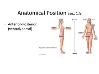
Stimuli—Orthopedic Anatomical Terminology. Anterior- in front of; ventral. Posterior. Posterior. Anterior. Anterior. Apophyses - a bony process or outgrowth that lacks an independent center of ossification like an epiphysis. Ischial tuberosity Ileac crest Greater trochanter

E N D
Anterior- in front of; ventral Posterior Posterior Anterior Anterior
Apophyses- a bony process or outgrowth that lacks an independent center of ossification like an epiphysis. Ischialtuberosity Ileac crest Greater trochanter Tibialtuberosity Base of 5thmetarsal
Base- the lower part or bottom of an object There is a Fx at the base of the 4th toe, proximal phalanx There is a Fx at the base of the proximal phalanx of the index finger, MTP jt.
Caudal- situated or oriented toward the tail end of an organism Cephalad- situated or oriented toward the head end of an organism or body part. There is a wedge compression Fx of the T 7 vertebra. In addition, there are a 2nd and 3rd more caudal, L1 and L2 compression fractures. Cephalad Caudal
Contralateral- on the opposite side There is a L hip fxcontralateral to the R femur fx
Coronal- a plane that divides the body into anterior and posterior parts The metal Foreign body (coin) is lying parallel to the coronal plane in an esophageal location at the T8 level.
Distal- situated away from the center of the body L wrist , distal ulna, metaphysealFx 5th toe, proximal phalanx, distal end Fx A distal fibula Fx
Dorsal- relating to the back or posterior i.e. the exterior part of the hand A Fx of the distal radius and ulna with 100% dorsal displacement of the distal fragments. Anterior Dorsal
Epiphysis- relating to a part of a long bone developed from a center of ossification distinct from the shaft(Often called a growth plate) Epiphysis Epiphysis Epiphysis
Frontal- relating to the anterior part of a body Posterior Posterior Frontal Frontal
Head- the rounded end of a bone Femoral Humeral Metacarpal Metatarsal Radial Fibular Comminuted Fx of the humeral head with signif displacement Fx of the 5th metacarpal head with minimal angulation Fx of the radial head , non displaced
Horizontal- a plane across the body at right angles to the coronal & sagittal planes resulting in upper and lower parts 375, 380, 391, 667
Inferior- below in relation to another structure; caudal (lower) There is a 3 part intertrochantericfx of the L hip. In addition , there are ipsilateral L superior and inferior pubic ramiFx s 1 3 2
Ipsilateral- on the same side There is a 3 part intertrochantericfx of the L hip. In addition , there are ipsilateral L superior and inferior pubic ramiFx s 1 3 2
Lateral- on the side; farther from the median or midsaggital plane There is a spiral fx of the lateral malleolus at the level of the mortise with approx 2 mm of displacement of the distal fragment. There is also widening of the medial joint mortise.
Longitudinal—aligned lengthwise; any plane perpendicular to the transverse plane
Medial- relating to the middle or center; nearer the median or midsagittal plane This is a bimallleolarfx of the L ankle. The medial malleolus has a transverse Fx that is non displaced. There is an old chip fx off the 1st toe, proximal phalanx, distal end , medial aspect . There is a new Fx of the 5th toe, prox phalanx which is angulated.
Metaphysis- relating to the growth zone between the epiphysis and the diaphysis during development of a bone
Neck- any constricted portion having a fancied resemblance to the neck of an animal Femoral Humeral Metacarpal Metatarsal Radial Fibular There is an oblique, minimally displaced fx of the neck of the 3rd metacarpal There is a fx of the R femoral neck.
Occipital- relating to the back of the head There is a fracture of the skull extending from the L temporal area to the high occiptal portion of the skull.
Palmar- volar; the flexor or anterior surface of the hand There is a Fx of the distal radius and ulna with palmar displacement of the distal fragments.
Posterior- behind or after in place There is an obvious posterior dislocation of the elbow present.
Proximal- nearest the trunk or the point of origin There is a non displaced transverse Fx of the prox 1/3 of the humerus There is an angulated transverse fx of the neck of the 5th proximal phalanx
Radial- relating to the lateral aspect of the upper limb There is a Fx of the L radial neck. . . The thumb is part of the radial aspect of the hand.
Rostral- situated at or directed toward the anterior (snout) end of any organism
Sagittal- a plane that goes from top to bottom dividing an object into a right and a left side. 1405, 1406 There is no sagittal deviation for this metal FB(coin) lodged in the distal 1/3 esophagus.
Superior- above in relation to another structure; cephalic, (higher) There are a superior and inferior pubic ramifx present in this view ofthepelvis.
Transverse- horizontal; lying across the body part in a horizontal plane. There is a displaced, angulated transverse fx of the neck of the proximal phalanx, 5th toe. There are non displaced transverse fxs of the diaphysis of the distal 1/3 radius and ulna.
Ulnar- relating to the medial aspect of the upper limb There is a intra-articularfx of the ulnar aspect of the 3rd finger, prox phalanx, at the MCP jt. There is an incomplete transverse fx of the distal ulnarstyloid.
Valgus- turned or bent outward There is a spiral fx of the R femur just distal to the hip prosthesis with a valgus displacement of the distal fragment There are fxs at the neck of the 2nd and 3rd metatarsals with a valgus deformity of the distal fragments
Varus- turned or bent inward There is a 3 part 100% displacement fx of the mid shaft L femur with varus displacement of the distal fragment.
Vertical- the direction aligned with the direction of he force of gravity There is an non displaced intra-articular vertical fx through the lateral tibial plateau of the knee • Left ileum—there is a non displaced vertical fx through the inferior aspect of the medial ileum There is a-non displaced, vertical fx through the L patella.
Volar- the palm of the hand or the sole of the foot There is a volar plate fx of the PIP jt, middle phalanx of the 2nd finger The volar aspect of the foot is same as the plantar aspect.