Download
1 / 23
230 likes | 493 Views
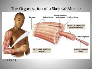
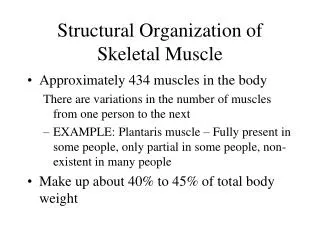
Organization of Skeletal Muscle. Skeletal Muscle Organization. Connective Tissue Coverings Skeletal muscles are attached to tendons which attach the muscle to bone Belly : Thicker part of muscle Epimysium : connective tissue layer that covers the muscle

E N D
Skeletal Muscle Organization • Connective Tissue Coverings • Skeletal muscles are attached to tendons which attach the muscle to bone • Belly: Thicker part of muscle • Epimysium: connective tissue layer that covers the muscle • Separates muscles from other structures/organs • Perimysium: Layer underneath the epimysium • Separates muscles into individual bundles • Endomysium: Layer underneath the perimysium • Surrounds each individual muscle fiber
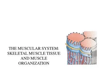
Structure of a Skeletal Muscle Fiber • Individual Muscle Fiber consists of: (outside to inside) • Outside of muscle fiber • Sarcolemma: cell membrane of muscle fiber • Sarcoplasm: cytoplasm of muscle fiber • Transverse Tubules or T-Tubules: connections from the sarcolemma to interior of muscle fiber for connection • Inside muscle fiber • Myofibrils: proteins that run the length of each fiber, surrounded by Sarcoplasmic Reticulum • S. R. stores Ca2+ ions which dictates muscle contraction
Structure of a Skeletal Muscle Fiber • Myofibril Components: • Several Sarcomeres made up of: • Actin: Thin Filaments, bundles with globular sections that fit into the binding sites on actin • Myosin: Thick Filaments, has binding sites to which actin attaches to during contraction
Muscle Contraction • T-tubules are stimulated from the nervous system • T-tubules release Ca2+ ions • The thin filaments/actin then are pulled along the thick filaments/myosin in place of where the Ca2+ ions were • http://www.youtube.com/watch?v=Ae0UYxqj3cM
Rigor Mortis • After death, ion pumps stop functioning calcium builds up in the sarcoplasm causing a fixed muscular contraction
Muscle Pain • Soreness after a workout • Stretching the muscle tissue farther than usual • Small microscopic tears to an individual muscle fiber • Skeletal muscle tissue is multinucleated + many neuromuscular junctions you mind sends signals when you are injured
Skeletal muscle identification • Muscular system can be divided into 2 groups: • Axial Muscles (60 %) • Appendicular Muscles (40%)
Neuromuscular Junction • Each skeletal muscle is controlled by a nerve cell called a motor neuron • Where the motor neuron and muscle fiber meet = Neuromuscular Junction
Axial Muscles • Muscles that position the head, spinal column, and move the rib cage. • Divided into 4 groups based on location and function: • Muscles of the head and neck • Muscles of the vertebral column • Oblique and rectus muscles • Muscles of the pelvic
Axial Muscles • Muscles of the Skull • Orbicularisoris • OrbicularisOculi • Nasalis • Zygomaticus Major • Zygomaticus Minor • Frontalis
Axial Muscles • Muscles of the Skull continued… • Platysma • Buccinator • Temporalis • Occipitalis • Masseter • Sternocleidomastoid
Axial Muscles • Muscles of the Back • Trapezius • LatissimusDorsi
Axial Muscles • Muscles of the Oblique • Pectoralis major • Serratus Anterior • Rectus Abdominis • External abdominal oblique
Appendicular Muscles • Muscles that support the pectoral and pelvic girdles, and the limbs. • Separated into 2 major groups: • Muscles of shoulders and upper limbs • Muscles of pelvis and lower limbs
Appendicular Muscles • Muscles of Pectoral girdle: • Trapezius • Rhomboid • Serratus anterior • Subcavius • Pectoralis minor
Appendicular Muscles • Muscles that move the arm: • Deltoid • Supraspinatus • Subscapularis • Infraspinatus • Coracobrachialis • Pectoralis major • Latissimusdorsi
Appendicular Muscles • Muscles of the arm: • Biceps brachii • Triceps brachii • Brachialis • Brachioradialis • Flexor carpiulnaris • Flexor carpidradialis • Palmaris longus • Extensor carpiradialis • Extensor carpiulnaris
Appendicular Muscles • Muscles of the Pelvic Girdle: • Gluteus maximus • Tensor fasciae latae • Gluteus medius • Gluteus minimus • Adductors • Iliopsoas
Appendicular Muscles • Muscles that move the lower limb: • Biceps femoris • Semimembranosus • Sartorius • Popliteus • Rectus femoris • semitendinosus
Appendicular Muscles • Muscles of the lower limb: • Gastrocnemius • Soleus • Fibularis • Tibialis posterior • Tibialis anterior • Extensor digitorumlongum • Calcaneal tendon