Download
1 / 26
260 likes | 295 Views
This article highlights the critical role of neuromuscular control in the rehabilitation process. Re-establishing neuromuscular control helps protect joints, prevent injuries, and complements traditional rehab methods. It focuses on the sensory feedback systems crucial for joint stability and proper muscle coordination. Key elements of neuromuscular control and motor patterns are discussed, along with the significance of muscle sense and proprioception. The text explores motor control mechanisms, including feed-forward and feed-back processes, and activities recommended for inducing neuromuscular adaptations. The physiology of mechanoreceptors, such as articular and musculotendon receptors, is explained to underscore their role in joint movement and proprioceptive feedback.

E N D
Why is it critical to the rehabilitation process? • Refocuses the athlete’s awareness of peripheral sensation & guides them into more coordinated motor strategies • Required to: • Protect joints from excessive strain • Provide prophylactic mechanism to recurrent injury • Complements traditional components of rehabilitation • We rely on sensory information from the periphery from our visual, vestibular, & somatosensory systems.
Primary role of articular structures • Stabilize & guide body segments • Provide mechanical restraint to abnormal joint motion • Dynamic restraint system • Capsuloligamentous tissue & musculotendon receptor sensory role • Detect joint motion & position • Detect changes in muscle length • Implicated in regulating muscle stiffness prior to loading • Injury results in damage to microscopic nerves associated with peripheral mechanoreceptors • Disrupts sensory feedback • Alters reflexive joint stabilization & neuromuscular coordination
Four critical elements of neuromuscular control in rehab • Joint sensation (position, motion, force) • Dynamic stability • Preparatory & reactive muscle characteristics • Conscious & unconscious functional motor patterns • Rehabilitation should address feedback systems • Preparatory (feed-forward) • Reactive (feed-back) • Muscle sense is divided into 4 sensory functions: • Sensation of passive movement • Sensation of active movement • Sensation of position • Sensations of heaviness & resistance
What is neuromuscular control? • Signal transmission through afferent sensory pathways • Proprioception • Conscious & unconscious appreciation of joint position • Awareness of position & movement • Any postural, positional or kinetic info provided to the CNS by sensory receptors in muscles, tendons or joints • Kinesthesia • Sensation of joint motion or acceleration • Sensation of ACTIVEmovement (contracting muscle) • Neuromuscular control • Efferent motor response to sensory information • Proprioception & kinesthesia
Motor control mechanisms • Feed-forward neuromuscular control • Planning movements based on sensory information from past experiences • Preparatory muscle activity • Operates on premise of initiating a motor response in anticipation of a load or activity • Feed-back neuromuscular control • Continuously regulates muscle activity through reflexive pathways • Reactive muscle activity • Operates directly in response to a potentially destabilizing event, using a normal reference point • Muscle stiffness • Ratio in change of force to change in length • Stiffer muscles resist stretching = more effective restraint to joint displacement • Modified by muscle activation
Activities for Inducing Adaptations • Open & closed kinetic chain activities • Balance training • Eccentric & high repetition low load exercises • Reflex facilitation • Stretch-shortening • Biofeedback training • Controlled positions of vulnerability
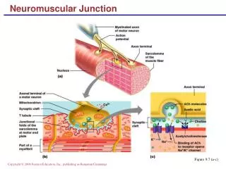
Physiology of Mechanoreceptors • Articular Mechanoreceptors • Specialized nerve endings that transduce mechanical tissue deformation into frequency modulated neural signals • Increased tissue deformation results in increased afferent firing rate or rise in quantity of mechanoreceptors activated • Types • Pacinian corpuscles – (Type II) sensitive to high-frequency vibration; compression sensitive • Ruffini endings – (Type I) sensitive to stretching of the joint capsule • Golgi-Mazzoni corpuscles – (Type III) sensitive to joint compression, not joint motion • Free nerve endings – (Type IV) stimulated by pain & inflammation when a joint is placed in an end position • Normally not active in normal joint movement
Articular Mechanoreceptors • Quick adapting (QA) • Cease discharging shortly after onset of stimulus • Provide conscious & unconscious kinesthetic sensation in response to joint movement/acceleration • Type II • Slow adapting (SA) • Continue to discharge as long as stimulus is present • Continuous feedback & proprioceptive information relative to joint position • Type I, III
Musculotendon Mechanoreceptors • Muscle spindles – located in the muscle • Responds to stretch of a muscle • Detects length & rate of length changes • Its stimulation leads to a contraction • Transmit information via afferent nerves • Innervated by small motor fibers (gamma efferents) • Project directly on motoneurons (monosynaptic reflexes) • Stretch reflex • Stimulation results in reflex contraction • Continued stimulation (gamma motor nerves) heighten stretch sensitivity • Muscle activity mediation
Musculotendon Mechanoreceptors • Golgi Tendon Organs (GTO) – located in tendon & musculotendon junction • Detects tension within a muscle & responds to both the contraction & stretching of a muscle • Regulate muscle activity & tension • Its stimulation results in muscle relaxation • GTO’s have opposite effect of muscle spindles by producing a relaxation in the muscle being loaded
Neural Pathways of Peripheral Afferents • Encoded signals - transmitted from peripheral receptors via afferent pathways (interneurons) to CNS • Brain Stem = Balance • Primary proprioceptive correlation center • Cerebral Cortex – location of conscious movement • Monosynaptic reflex pathway - links muscle spindles directly to motor nerves • Balance • Influenced by peripheral afferent mechanism mediating joint proprioception • Partially dependent on inherent ability to integrate joint position sense, vision & vestibular apparatus with neuromuscular control
Re-establishing Neuromuscular Control • Injuries result in decreases in neuromuscular control • Pathoetiology • Injury results in deafferentation of ligament & capsular mechanoreceptors • Joint inflammation & pain compound sensory deficits • Congenital/pathological joint laxity have diminished ability to detect joint motion & position • Proprioceptive, kinesthetic deficits & mechanical instability lead to functional instability
Objectives for Neuromuscular Rehabilitation • Develop/re-establish afferent & efferent characteristics that enhance dynamic stability • Elements • Proprioceptive & kinesthetic sensation • Dynamic joint stabilization • Reactive neuromuscular control • Functional motor patterns • Afferent & Efferent Characteristics • Sensitivity of peripheral receptors • Facilitation of afferent pathways • Muscle stiffness • Onset rate & magnitude of muscle activity • Simultaneous activation of agonist/antagonist • Reflexive & discriminatory muscle activation
Neuromuscular Characteristics • Peripheral Afferent Receptors • Altered peripheral afferent information may disrupt motor control & functional stability • Repetitious athletic activity enhances proprioceptive & kinesthetic acuity = facilitated afferent pathways • Enhanced joint motion awareness improves feed-forward & feedback mechanisms • Muscle Stiffness • Significant role in preparatory & reactive dynamic restraints • Exercises that encourage muscle stiffness should be incorporated into rehabilitation programs • Eccentric exercises • Chronic overload results in connective tissue proliferation, desensitizing GTO’s & increase muscle spindle activity • Power trained vs. Endurance trained athletes • Power athlete = Faster muscle pre-activation (EMG) • Endurance athlete = Increased baseline motor tone
Reflexive Muscle Activation • Reflex latency times may be dependent on types of training (endurance vs. power) • Preparatory & reactive muscle activation might improve dynamic stability & function if muscle stiffness is enhanced in deficient joints • Decreasing electromechanical delay between joint loading & protective muscle activation can increase stability & function
Discriminate Muscle Activation • Unconscious control of muscle activity is critical in balance & coordination • May initially require conscious activation prior to unconscious control • Use of biofeedback can aid in this process • Help eliminate imbalances & re-establish preparatory & reactive muscle activity
Elements for Neuromuscular Control • Proprioception & Kinesthesia Training • Restore neurosensory properties • Enhance sensitivity of uninvolved peripheral afferents • Joint compression is believed to maximally stimulate articular receptors • Closed chain exercises through available ROM • Early repositioning tasks are critical • Conscious to unconscious joint awareness • Applying neoprene sleeve or ace wrap stimulates cutaneous receptors – additional proprioception & kinesthesia
Dynamic Stabilization • Encourage preparatory agonist/antagonist coactivation • Restores force couples & balances joint forces • Results in decreased loads on static structures • Activities that require anticipatory & reactive adjustments to imposed loads • Combination of balance & stretch shortening exercises • Encourages preparatory & reactive muscle activity • Closed chain exercises induce coactivation & compression
Reactive Neuromuscular Control • Stimulates reflex pathways • Object is to impose perturbations that stimulate reflex stabilization • Can resultin decreased response time & develop reactive strategies to unexpected joint loads • Perturbations should be unexpected in order to facilitate reflexive activity • Functional Activities • Objective is to return athlete to pre-injury activity • Involves sports specific movement patterns designed to restore functional ability • Can be utilized to assess readiness for return to play • Stresses peripheral afferents, simultaneous muscle activation, reflexive activity • Progress from conscious to unconscious • Develop functionally specific movement patterns, ultimately decreasing risk of injury
Lower Extremity Techniques • Techniques should focus on muscle groups that require attention • Progress from no weight to weight assisted • Use of closed-chain activities is encouraged • Replicates environmental demands • Plays on principles of neuromuscular control • Joint stabilization exercises • Balance & partial weight bearing activities • Progress non-weight bearing to full weight-bearing • Balance on unstable surfaces can begin once full-weight bearing
Slide board exercises • Stimulates coactivation with increasing muscle force & endurance • Stimulating dynamic stability & stiffness • Stair climbing (forward & backward) • Emphasis on eccentric strength • Biofeedback • Used to develop agonist/antagonist coactivation • Encourages voluntary muscle activation • Stretch-shortening exercises • Eccentric deceleration & explosive concentric contractions • Incorporate early in process (modified loads) • Involves preparatory & reactive muscle activity • Hopping progression • Double Single leg • Sagittal Lateral Rotational hopping • Surface modification
Rhythmic stabilization React to joint perturbations preparatory & reactive muscle activity Alterations in loads & displacement Unstable surfaces Linear & angular perturbations, altering center of gravity Facilitate reflexive activity Ball toss Disrupt concentration, induce unconscious response & reactive adaptation Trampoline Hopping Hopping & landing (double support, single support, rotation) Challenge athlete Hopping & catching Hopping & landing on varying surfaces Functional activities Restore normal gait Athlete must internalize normal kinematics (swing & stance) Utilize retro walking (hamstring activity), pool or unloading devices Cross over walking, figure 8’s, cutting, carioca, changes in speed Functional activities that simulate demands of sport
Upper Extremity Techniques • Work to maintain joint congruency & functional stability • Requires dynamic restraint via coordinated muscle activation • Injury to static stabilizers • Failure of dynamic restraint system • Could result in repetitive loads, compromising joint integrity & predisposing athlete to re-injury • Adapt lower extremity exercise for upper extremity
Muscle stiffness • Enhance using elastic resistance (focus on eccentrics) • High repetitions & low resistance • Upper extremity ergometers should be incorporated for endurance • Dynamic stabilization • Stability platforms • Push-ups, horizontal abduction, tracing circles on slide board with dominant & non-dominant arms • Plyometric exercise
Reactive Neuromuscular Exercises • Manual perturbations • Rhythmic stabilization with gradual progression • Placing joint in inherently unstable positions • Functional Training • Developing motor patterns in overhead position • Reproduce demands of activity • Emphasis on technique • Re-education of functional patterns • Speed & complexity in movement require rapid integration of sensory information