Download
1 / 38
390 likes | 459 Views
Learn about blood flow types and characterizations, flow variations, resistance, and Doppler shifts in a concise guide.

E N D
Display of Motion &Doppler Ultrasound • Resident Class
Hemodynamics • Plug • Laminar • Disturbed • Turbulent Blood Flow Characterization
Plug Flow • Type of normal flow • Constant fluid speed across tube • Occurs near entrance of flow into tube
Laminar Flow • also called parabolic flow • fluid layers slide over one another • occurs further from entrance to tube • central portion of fluid moves at maximum speed • flow near vessel wall hardly moves at all • friction with wall
Flow • Disturbed Flow • Normal parallel stream lines disturbed • primarily forward particles still flow • Turbulent Flow • random & chaotic • individual particles flow in all directions • net flow is forward • Often occurs beyond obstructionsuch as plaque on vessel wall
Flow, Pressure & Resistance • Pressure • pressure difference between ends of tube drives fluid flow • Resistance • more resistance = lower flow rate • resistance affected by • fluid’s viscosity • vessel length • vessel diameter • flow for a given pressure determined by resistance
Flow Variations • pressure & flow in arteries fluctuate with pulse • pressure & flow in veins much more constant • pulse variations dampened by arterial system
Flow Rate Measurements • Volume flow rate • Volume of liquid passing a point per unit time • Example • 100 ml / second
Flow Rate Measurements • Linear flow rate • Distance liquid moves past a point per unit time • Example • 10 cm / second
Flow Rate Measurements Volume Flow Rate = Linear flow rate X Cross Sectional Area
Flow Rate Measurements Volume Flow Rate = Linear flow rate X Cross-sectional Area High Velocity Small Cross-section Low Velocity Large Cross-section Same Volume Flow Rate
Volume Flow Rates • constant volume flow rate in all parts of closed system Sure! Any change in flow rate would mean you’re gaining or losing fluid.
Stenosis • narrowing in a vessel • fluid must speed up in stenosis to maintain constant flow volume • no net gain or loss of flow • turbulent flow common downstream of stenosis
Stenosis • If narrowing is short in length • Little increase in overall resistance to flow • Little effect on volume flow rate • If narrowing is long • Resistance to flow increased • Volume flow rate decreased
Doppler Shift • difference between received & transmitted frequency • caused by relative motion between sound source & receiver • Frequency shift indicative of reflector speed OUT IN
Doppler Examples • change in pitch of as object approaches & leaves observer • train • Ambulance siren • moving blood cells • motion can be presented as sound or as an image
q Doppler Angle • angle between sound travel & flow • 0 degrees • flow in direction of sound travel • 90 degrees • flow perpendicular to sound travel
Flow Components • Flow vector can be separated into two vectors Flow parallel to sound Flow perpendicular to sound
Doppler Sensing • Only flow parallel to sound sensed by scanner!!! Flow parallel to sound Flow perpendicular to sound
Doppler Sensing • Sensed flow always < actual flow Actual flow Sensed flow
Doppler Sensing • cos(q) = SF / AF Actual flow (AF) q Sensed flow (SF) q
Doppler Equation 2 X fo X v X cosq f D = fe - fo = ------------------------- c • where fD =Doppler Shift in MHz fe = echo of reflected frequency (MHz) fo = operating frequency (MHz) v = reflector speed (m/s) q = angle between flow & sound propagation c = speed of sound in soft tissue (m/s) q
Relationships 2 X fo X v X cosq f D = fe - fo = ------------------------- c • positive shift when reflector moving toward transducer • echoed frequency > operating frequency • negative shift when reflector moving away from transducer • echoed frequency < operating frequency q q
cosq q Relationships 2 X fo X v X cosq f D = fe - fo = ------------------------- c • Doppler angle affects measured Doppler shift q
Simplified (?) Equation 2 X fo X v X cosq f D = fe - fo = ------------------------- c • Solve for reflector velocity • Insert speed of sound for soft tissue • Stick in some units 77 X fD (kHz) v (cm/s) = -------------------------- fo (MHz) X cosq Simplified:
Doppler Relationships • higher reflector speed results in greater Doppler shift • higher operating frequency results in greater Doppler shift • larger Doppler angle results in lower Doppler shift 77 X fD (kHz) v (cm/s) = -------------------------- fo (MHz) X cos
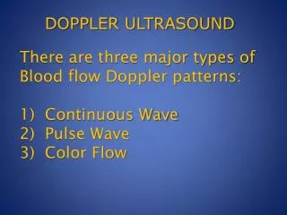
Continuous Wave Doppler • Audio presentation only • No image • Useful as fetal dose monitor
Continuous Wave Doppler • 2 transducers used • one continuously transmits • voltage frequency = transducer’s operating frequency • typically 2-10 MHz • one continuously receives • Reception Area • flow detected within overlap of transmit & receive sound beams
- = Continuous Wave Doppler:Receiver Function • receives reflected sound waves • Subtract signals • detects frequency shift • typical shift ~ 1/1000 th of source frequency • usually in audible sound range • Amplify subtracted signal • Play directly on speaker
Doppler Pulses • short pulses required for imaging • minimizes spatial pulse length • optimizes axial resolution • longer pulses required for Doppler analysis • reduces bandwidth • provide purer transmitted frequency • important for accurate measurement of frequency differences needed to calculate speed
Color-Flow Display Features • Imaged electronically scanned twice • imaging scan processes echo intensity • Doppler scan calculates Doppler shifts • Reduced frame rates • only 1 pulse required for imaging • additional pulses required when multiple focuses used • several pulses may be required along a scan line to determine Doppler shift
Gate Duplex Doppler Gates • operator indicates active Doppler region on display • regions are called gates • only sound in gate analyzed for frequency shift • can be isolated based on delay time after pulse
Spectral Display • shows range of frequencies received • amplitude of each frequency indicated by gray shade • can be displayed real time • fast Fourier Transform (FFT) technique frequency range Frequency Elapsed Time
Spectral Broadening • display indicates range of frequencies • corresponds to range of speeds of blood cells • range indicative of type of flow • laminar, disturbed, turbulent frequency range Frequency Time
Pulse Wave Doppler • Allows range selectivity • monitor Doppler shift (frequency difference) at only selected depth(s) • ability to separate flow from >1 vessel or localize flow within vessel
Spectral vs. Color-Flow • spectral Display shows frequency range directly • Color Doppler’s color represents complete spectrum at each pixel frequency range Frequency Elapsed Time
Power Doppler • AKA • Energy Doppler • Amplitude Doppler • Doppler angiography • Magnitude of color flow output displayed rather than Doppler frequency signal • flow direction or different velocities not displayed "Color Power Angio" of the Circle of Willis