Download
1 / 58
610 likes | 650 Views
Dive deep into the intricate world of chromatin and chromosomes, exploring euchromatin, heterochromatin, banding patterns, centromeres, and more crucial biological concepts.

E N D
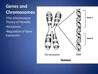
6.2 Chromatin is divided into euchromatin and heterochromatin • Individual chromosomes can be seen only during mitosis. • During interphase, the general mass of chromatin is in the form of euchromatin. • Euchromatin is less tightly packed than mitotic chromosomes. • Regions of heterochromatin remain densely packed throughout interphase.
6.3 Chromosomes have banding patterns • Certain staining techniques cause the chromosomes to have the appearance of a series of striations called G-bands. • The bands are lower in G • C content than the interbands. • Genes are concentrated in the G • C-rich interbands.
6.4 Eukaryotic DNA has loops and domains attached to a scaffold • DNA of interphase chromatin is negatively supercoiled into independent domains of ~85 kb. • Metaphase chromosomes have a protein scaffold to which the loops of supercoiled DNA are attached.
6.5 Specific sequences attach DNA to an interphase matrix • DNA is attached to the nuclear matrix at specific sequences called MARs or SARs. • The MARs are A • T-rich but do not have any specific consensus sequence.
6.6 The centromere is essential for segregation • A eukaryotic chromosome is held on the mitotic spindle by the attachment of microtubules to the kinetochore that forms in its centromeric region. • Centromeres often have heterochromatin that is rich in satellite DNA sequences.
6.7 Centromeres have short DNA sequences in S. cerevisiae • CEN elements are identified in S. cerevisiae by the ability to allow a plasmid to segregate accurately at mitosis. • CEN elements consist of short conserved sequences CDE-I and CDE-III that flank the A • T-rich region CDE-II.
6.8 The centromere binds a protein complex • A specialized protein complex that is an alternative to the usual chromatin structure is formed at CDE-II. • The CBF3 protein complex that binds to CDE-III is essential for centromeric function. • The proteins that connect these two complexes may provide the connection to microtubules.
The CEN region recruits three DNA-binding factors: Cbfl, CBF3 (an essential four-protein complex ), and Mif2 (CENP-C in multicellular eukaryotes). In addition, a specialized chromatin structure is built by binding the CDE-ll region to a protein called Cse4, a histone H3 variant (analogous to CENP-A in multicellular eukaryotes), probably in the context of an otherwise nonnal nucleosome. A protein called Scm3 is required for proper association of Cse4 with GEN. Inclusion of CenH3 histone variants related to Cse4 are a universal aspect of centromere construction in all species. The basic interaction consists of bending the DNA of the CD E-ll region around a protein aggregate; thereaction is probably assisted by the occurrence of intrinsic bending in the CDE·lf sequence.CDE-1 is bound by a bomodimer of Cbfl; this interaction i s not essential for centromere function, but in its absence the fidelity of chromosome segregation is reduced - 1OX. The 240-kD heterotetramer, CBF3, binds to CDE-111. This interaction is essential for centromeric function.The proteins bound at CDE-1, CDE-Il, and CD Ell/also interact wi th another group of proteins(Ctfl9, Mm12 1, and Okpl}, which in turn link the centromeric complex to the kinetochore proteins ( -70 individual kinetochore proteins have been identi6ed in yeast) ;md to the microtubule.
6.9 Centromeres may contain repetitious DNA • Centromeres in higher eukaryotic chromosomes contain large amounts of repetitious DNA. • The function of the repetitious DNA is not known.
6.10 Telomeres are replicated by a special mechanism • The telomere is required for the stability of the chromosome end. • A telomere consists of a simple repeat where a C+A-rich strand has the sequence C>1(A/T)1-4.
6.11 Telomeres seal the chromosome ends • The protein TRF2 catalyzes a reaction in which: • the 3repeating unit of the G+T-rich strand forms a loop by displacing its homologue in an upstream region of the telomere.
6.12 Lampbrush chromosomes are extended • Sites of gene expression on lampbrush chromosomes show loops that are extended from the chromosomal axis.
6.13 Polytene chromosomes form bands • Polytene chromosomes of Dipterans have a series of bands that can be used as a cytological map.
6.14 Polytene chromosomes expand at sites of gene expression • Bands that are sites of gene expression on polytene chromosomes expand to give “puffs.”
6.15 The nucleosome is the subunit of all chromatin • Micrococcal nuclease releases individual nucleosomes from chromatin as 11S particles. • A nucleosome contains: • ~200 bp of DNA • two copies of each core histone (H2A, H2B, H3, and H4) • one copy of H1 • DNA is wrapped around the outside surface of the protein octamer.
6.16 DNA is coiled in arrays of nucleosomes • Greater than 95% of the DNA is recovered in nucleosomes or multimers when micrococcal nuclease cleaves DNA of chromatin. • The length of DNA per nucleosome varies for individual tissues in a range from 154-260 bp.
6.17 Nucleosomes have a common structure • Nucleosomal DNA is divided into the core DNA and linker DNA depending on its susceptibility to micrococcal nuclease. • The core DNA is the length of 146 bp that is found on the core particles produced by prolonged digestion with micrococcal nuclease.
6.17 Nucleosomes have a common structure • Linker DNA is the region of 8-114 bp that is susceptible to early cleavage by the enzyme. • Changes in the length of linker DNA account for the variation in total length of nucleosomal DNA. • H1 is associated with linker DNA and may lie at the point where DNA enters and leaves the nucleosome.
6.18 DNA structure varies on the nucleosomal surface • 1.65 turns of DNA are wound around the histone octamer. • The structure of the DNA is altered so that it has: • an increased number of base pairs/turn in the middle • but a decreased number at the ends
6.18 DNA structure varies on the nucleosomal surface • Approximately 0.6 negative turns of DNA are absorbed by the change in bp/turn from 10.5 in solution to an average of 10.2 on the nucleosomal surface. • This explains the linking number paradox.
6.19 Organization of the histone octamer • The histone octamer has a kernel of a H32 • H42 tetramer associated with two H2A • H2B dimers. • Each histone is extensively interdigitated with its partner.
6.19 Organization of the histone octamer • All core histones have the structural motif of the histone fold. • The histone N-terminal tails extend out of the nucleosome.
6.20 The path of nucleosomes in the chromatin fiber • 10-nm chromatin fibers are unfolded from 30-nm fibers and consist of a string of nucleosomes. • 30-nm fibers have 6 nucleosomes/turn, organized into a solenoid. • Histone H1 is required for formation of the 30-nm fiber.
6.21 Reproduction of chromatin requires assembly of nucleosomes • Histone octamers are not conserved during replication; • However, H2A • H2B dimers and H32 • H42 tetramers are conserved. • There are different pathways for the assembly of nucleosomes during replication and independently of replication. • Accessory proteins are required to assist the assembly of nucleosomes.
6.21 Reproduction of chromatin requires assembly of nucleosomes • CAF-1 is an assembly protein that is linked to the PCNA subunit of the replisome; • it is required for deposition of H32 • H42 tetramers following replication. • A different assembly protein and a variant of histone H3 may be used for replication-independent assembly.
6.22 Do nucleosomes lie at specific positions? • Nucleosomes may form at specific positions as the result either of: • the local structure of DNA • proteins that interact with specific sequences • The most common cause of nucleosome positioning is when proteins binding to DNA establish a boundary. • Positioning may affect which regions of DNA are in the linker and which face of DNA is exposed on the nucleosome surface.
6.23 Domains define regions that contain active genes • A domain containing a transcribed gene is defined by increased sensitivity to degradation by DNAase I.
6.24 Are transcribed genes organized in nucleosomes? • Nucleosomes are found at the same frequency when transcribed genes or nontranscribed genes are digested with micrococcal nuclease. • Some heavily transcribed genes appear to be exceptional cases that are devoid of nucleosomes.
6.25 Histone octamers are displaced by transcription • RNA polymerase displaces histone octamers during transcription in a model system; • Octamers reassociate with DNA as soon as the polymerase has passed. • Nucleosomes are reorganized when transcription passes through a gene.
6.26 Nucleosome displacement and reassembly require special factors • Ancillary factors are required both: • for RNA polymerase to displace octamers during transcription • for the histones to reassemble into nucleosomes after transcription
6.27 DNAase hypersensitive sites change chromatin structure • Hypersensitive sites are found at the promoters of expressed genes. • They are generated by the binding of transcription factors that displace histone octamers.
6.28 Chromatin remodeling is an active process • Chromatin structure is changed by remodeling complexes that use energy provided by hydrolysis of ATP. • The SWI/SNF, RSC, and NURF complexes all are very large; • there are some common subunits.
6.28 Chromatin remodeling is an active process • A remodeling complex does not itself have specificity for any particular target site; • it must be recruited by a component of the transcription apparatus. • Remodeling complexes are recruited to promoters by sequence-specific activators. • The factor may be released once the remodeling complex has bound.
6.19 Histone acetylation is associated with genetic activity • Histone acetylation occurs transiently at replication. • Histone acetylation is associated with activation of gene expression. • Deacetylated chromatin may have a more condensed structure.
6.19 Histone acetylation is associated with genetic activity • Transcription activators are associated with histone acetylase activities in large complexes. • The remodeling complex may recruit the acetylating complex. • Histone acetylases vary in their target specificity.
6.19 Histone acetylation is associated with genetic activity • Acetylation could affect transcription in a quantitative or qualitative way. • Deacetylation is associated with repression of gene activity.
6.19 Histone acetylation is associated with genetic activity • Deacetylases are present in complexes with repressor activity. • Acetylation of histones may be the event that maintains the complex in the activated state.
6.30 Heterochromatin propagates from a nucleation event • Heterochromatin is nucleated at a specific sequence. • The inactive structure propagates along the chromatin fiber. • Genes within regions of heterochromatin are inactivated.
6.30 Heterochromatin propagates from a nucleation event • The length of the inactive region varies from cell to cell. • Inactivation of genes in this vicinity causes position effect variegation. • Similar spreading effects occur at: • telomeres • the silent cassettes in yeast mating type