Download
1 / 78
830 likes | 1.17k Views
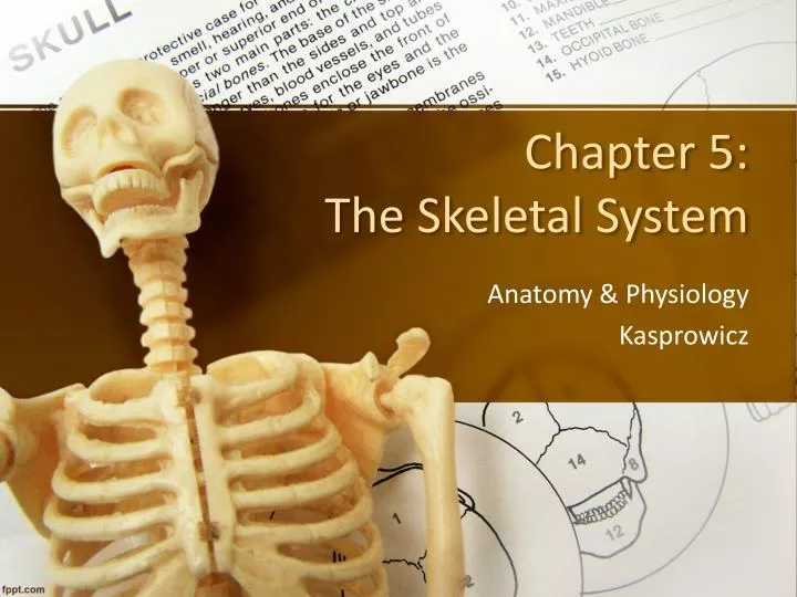
Chapter 5: The Skeletal System . Anatomy & Physiology Kasprowicz. Parts of the Skeletal System. Bones (make up the skeleton) Joints (articulations) Cartilages Ligaments (dense, regular connective tissue connecting bone to bone). Divisions of the Skeleton. Axial Skeleton

E N D
Chapter 5: The Skeletal System Anatomy & Physiology Kasprowicz
Parts of the Skeletal System • Bones (make up the skeleton) • Joints (articulations) • Cartilages • Ligaments (dense, regular connective tissue connecting bone to bone)
Divisions of the Skeleton Axial Skeleton • bones that form the longitudinal axis • skull, vertebrae, rib cage Appendicular Skeleton • bones of the limbs (appendages) & girdles • arms, legs, pelvis
Functions of the Skeleton • Support for the body our internal framework • Protection of soft organs ex. skull brain rib cage lungs & heart vertebrae spinal cord
Functions of the Skeleton • Movement skeletal muscles use the bones as levers to move the body & its parts • Storage Bone tissue stores minerals, such as calcium and phosphorus Fat is stored in yellow marrow
Functions of the Skeleton 5) Blood Cell Formation Hematopoiesis occurs in red marrow The adult skeleton is composed of 206 bones!
Classification of Bones Bones can be classified based on their SHAPE or HISTOLOGICAL ORGANIZATION (STRUCTURE)
Classification of Bones: Shape • Long bones • Typically long & slender • Shaft with heads at both ends • Mostly compact bone Examples: arms, legs, fingers, toes
Classification of Bones: Shape 2) Short bones • Typically box- or cube-shaped • Mostly spongy bone Examples: wrists & ankles
Classification of Bones: Shape 3) Flat bones • thin, flattened • usually curved • thin layers of compact bone surrounding spongy bone ribs
Classification of Bones: Shape 4) Irregular bones • Do not fit in other categories Examples: Vertebrae Hip bones
Types of Bone (Osseous) Tissue All bones contain both types of bone tissue. Their relationship & proportions vary depending on bone shape. Compact Bone • dense, smooth & homogenous Spongy Bone • open network of struts & plates
Gross Anatomy of a Long Bone • diaphysis a) tubular shaft b) compact bone • epiphysis a) ends of the bone b) mostly spongy bone
Structures of a Long Bone • periosteum a) outside covering of the diaphysis b) fibrous connective tissue membrane • Sharpey’s fibers connect periosteum to the underlying bone
Structures of a Long Bone 3) Arteries supply the osteocytes w/ nutrients & oxygen • Articular Cartilage a) covers the epiphyses b) made of hyaline cartilage c) decreases friction at the joints
Structures of a Long Bone • Medullary Cavity a) Cavity of the shaft b) bone marrow consists of loose connective tissue - yellow marrow (fat storage) in adults - Contains red marrow (for blood cell formation) in infants c) endosteum layer of cells lining the cavity
Bone Histology • Osseous tissue (bone) is a type of connective tissue • Four Characteristics of Bone 1) bone matrix is very dense & contains calcium salts 2) contains bone cells (osteocytes) in pockets called lacunae
Bone Histology Four Characteristics of Bone cont… 3) Canaliculi allow for gas exchange and diffusion of nutrients and waste products 4) except at joints, bones are covered by the a periosteum Let’s take a look at each characteristic…
Bone Histology: Matrix • 2/3 of bone matrix is calcium phosphate, Ca3(PO4)2 • w/ calcium hydroxide, forms crystals of hydroxyapatite, which incorporate other calcium salts & minerals into the matrix • 1/3 of bone matrix is collagen fibers Only 2% of bone mass comes from cells
Bone Histology: Cells • There are 4 types of bone cells osteocytes are the most common • Osteocytes sit in a pocket-like structure called a lacuna (lacunae, plural) • Lacunae are arranged in concentric rings, or layers of matrix, called lamellae (lamella, singular)
Bone Histology: Canaliculi Canaliculi (tiny canals) • Radiate from the central canal (blood vessels) to lacunae • Form a transport system between osteocytes and between the central canal & osteocytes
Bone Histology: Periosteum • Covers all bones, except parts enclosed in joint capsules 2) It is made up of an: • outer, fibrous layer • inner, cellular layer
Bone Histology: Periosteum • Functions of periosteum a) Isolate bone from surrounding tissues b) Provide a route for circulatory and nervous supply c) Participate in bone growth and repair
Compact Bone • Osteon (Haversian System) osteon – the basic unit of compact bone • Organization of an Osteon a) Osteocytes are arranged in concentric lamellae around a central (Haversian) canal containing blood vessels
Compact Bone • Organization of an Osteon cont… b) Central (Haversian) canal opening in the center of an osteon containing blood vessels and nerves c) Perforating (Volkman’s) canal perpendicular to the central canal also containing blood vessels and nerves
Compact Bone osteon
Spongy Bone 1) Does not have osteons • A “honeycomb” matrix forms an open network of trabeculae • Trabeculae have no blood vessels
Spongy Bone: Yellow Marrow In some bones, spongy bone holds yellow bone marrow which is used to store fat
Spongy Bone: Red Marrow The space between trabeculae is filled with red bone marrow. Red marrow forms red blood cells and supplies nutrients to osteocytes.
Bone Markings • Surface features of bones that show… 1) Sites of attachment for muscles, tendons, and ligaments 2) Passages for nerves and blood vessels Categories of bone markings • projections or processes– grow out from the bone surface • depressionsor cavities – indentations
The structure of bone is specialized for flexibility and tensile strength. Bone Formation, Growth & Remodeling
Bone Formation • In embryos, the skeleton is primarily hyaline cartilage • During fetal development, much of this cartilage is replaced by bone • By the end of the toddler years, cartilage remains only in isolated areas: - bridge of the nose - parts of ribs - articular cartilage (in the joints) - epiphyseal plates
Bone Formation • The process of “bone formation” is called ossification. 1) Unless it is a flat bone, a hyaline cartilage model is covered with bone matrix by osteoblasts. osteoblasts- immature, bone-forming cells Over time, the “cartilage bone” is enclosed in a layer of real bone (kind of like an arm in a cast).
Bone Formation • The enclosed layer of hyaline cartilage is digested away by osteoclasts. osteoclasts- bone destroying cells; break down bone matrix for remodeling and release of calcium The breakdown of the cartilage “model” leaves a medullary cavity in the bone.
Bone Growth Epiphyseal plates allow for growth in the length of long bone during childhood & adolescence