Download
1 / 12
0 likes | 18 Views
This study evaluated the effect of lime juice extract of cashew bark (LJECB) on serum antioxidant status in normal and Wistar rats with indomethacin toxicity. Fresh Anacardium occidentale bark was pulverised and extracted using fresh juice of Citrus aurantifolia. Forty two female Wistar rats (150-200 g) were purchased and randomised into seven groups (n=6): Group 1 (normal control); Group 2 (Indomethacin 30 mg/kg only); Group 3 (Indomethacin 30 mg/kg Gecrol antacid); Group 4 (Indomethacin 30 mg/kg LJECB 400 mg/kg); Group 5 (Indomethacin 30 mg/kg LJECB 800mg/kg);

E N D
26 Int. J. Biomol. Biomed. International Journal of Biomolecules and Biomedicine (IJBB) ISSN: 2221-1063 (Print), 2222-503X (Online) http://www.innspub.net Vol. 18, No. 1, p. 26-37, 2024 RESEARCH PAPER RESEARCH PAPER RESEARCH PAPER RESEARCH PAPER OPEN ACCESS OPEN ACCESS OPEN ACCESS OPEN ACCESS Evaluation of antioxidant activity of lime juice extract of cashew bark in normal and wistar rats with indomethacin toxicity Godwin Anyim*, Morakinyo Adetoun Elizabeth, Babarinde Samuel Olufolarin, Bamgboye Faith Funke, Akinbinu Ibukunoluwa Rebecca, Oyedemi Temitope, Oyedepo Temitope Adenike Department of Biochemistry, Faculty of Science, Adeleke University, Ede, Osun State, Nigeria Key words:Indomethacin toxicity, Antioxidant, Anacardium occidentale, Citrus aurantifolia Article Published: 21 February 2024 Abstract This study evaluated the effect of lime juice extract of cashew bark (LJECB) on serum antioxidant status in normal and Wistar rats with indomethacin toxicity. Fresh Anacardium occidentale bark was pulverised and extracted using fresh juice of Citrus aurantifolia. Forty two female Wistar rats (150-200 g) were purchased and randomised into seven groups (n=6): Group 1 (normal control); Group 2 (Indomethacin 30 mg/kg only); Group 3 (Indomethacin 30 mg/kg + Gecrol antacid); Group 4 (Indomethacin 30 mg/kg + LJECB 400 mg/kg); Group 5 (Indomethacin 30 mg/kg + LJECB 800mg/kg); Group 6 (LJECB 400 mg/kg only), Group 7 (LJECB 800 mg/kg only). Post-treatment with LJECB or Gecrol antacid was commenced 4 hours for 14 days. On the 15th day, the animals were sacrificed, and the serum was collected aand used for antioxidant, lipid peroxidation (malonialdehyde MDA), and total protein assays based on standard protocols. Results showed that treatment with 400 mg/kg LJECB elevated the nitric oxide (NO), superoxide dismutase (SOD), and MDA levels, but lowered the activities of glutathione peroxidase (GPx) and catalase (CAT). Also, 800 mg/kg of LJECB elevated the NO, CAT, SOD, and MDA levels, but lowered the level of GSH, GPx, and total protein. The study concludes that LJECB provoked a significant increase in antioxidant status (NO, SOD, and CAT) but lowered the activity of GPx and reduced glutathione (GSH). However, the elevation of lipid peroxidation biomarker (MDA) suggests a possible damage to the stomach endothelial membranes. Therefore, caution should be exercised when consuming the LJECB for therapy. *Corresponding Author: Godwin Anyim godwin.anyim@adelekeuniversity.edu.ng Anyim et al.
27 Int. J. Biomol. Biomed. Introduction (DNA), and prevent the production of energy (Sharifi- Oxidative stress has been described as a component Rad et al., 2020). of many disease conditions (Forman and Zhang, 2021). Oxidative stress is a physiological condition Increased production of ROS is a hallmark that characterized by an imbalance between oxidant characterises the toxicity of many xenobiotics, production and the body's antioxidant defense including chemotherapeutic drugs (Jomova et al., mechanisms in favour of oxidants, leading to damage 2023). The impact of oxidative stress on a biological in biological systems (Aminjan et al., 2019). Free system is dependent on several factors including the radicals including reactive oxygen species (ROS) and nature of oxidant, the site, and intensity of peroxides are known to mediate cellular damage via production, a functional repair system, and oxidation of biomolecules including lipids, proteins, endogenous antioxidant system (Pizzino et al., 2017). and DNA (Ayala et al., 2014). Oxidative stress has Since the organism may not be able to control these been linked to the development of several chronic factors on its own, there is a need to fortify the body conditions including cardiovascular diseases, with abundant antioxidant compounds to enable it neurodegenerative disorders, premature aging, DNA protect the body from free radical insult; irrespective disruption, lipid peroxidation, and gastric mucosal of the source, type, and abundance (Fountoucidou et injuries (Valko et al., 2007). The extent to which al., 2019). oxidants promote human diseases varies in biological systems. Therefore, raising the antioxidant status may Antioxidants, whether in form of diets or not be effective in all disease conditions (Forman and supplements, play a crucial role in maintaining Zhang, 2021). The context of oxidants or reactive cellular health by neutralizing ROS and preventing species is comprised of oxygen and nitrogen radicals, oxidative damage (Vona et al., 2021). Antioxidants each with unique reactive properties and interactions can be obtained from various sources including fruits, (Murphy et al., 2022). Therefore, there is a need to seeds, vegetables, and herbs (Saleem et al., 2023). study the species of the oxidants that promote cellular Citrus fruits are particularly known for their rich damage to provide a holistic approach in rational antioxidant content due to the presence of bioactive antioxidant drug design. compounds like ascorbic acid, phenolic compounds, and flavonoids (Saleem et al., 2023). Lime (Citrus Several studies have evaluated oxidative stress in aurantifolia) is one such citrus fruit that has gained living systems and observed that production of ROS is attention for its potential health benefits (Rahaman et dependent on enzymatic and non-enzymatic reactions al., 2023). Synthetic antioxidants are known to (Pizzino et al., 2017). The human body produces the protect against oxidative damages. However, research oxidants or free radicals via the normal biological has shown that some synthetic antioxidants are not as processes, including breathing, food digestion, effective as natural antioxidants as they produce toxic alcohol or drug metabolism, and energy production. effects when consumed as supplements or remedies The natural antioxidant mechanism normally helps to (Sen, 2014). eliminate the free radicals. A controlled production of free radicals were reported to have beneficial roles in The genus Anarcadium has received great recognition cellular defense against invasive pathogens, cell in recent years due to their nutritional, biological, and signaling, etc (Saleem et al., 2023). However, if the pharmacological benefits (Salehi et al., 2020). The body fails to control the production or scavenge them, leaves, fruits, and stem parts contain beneficial they can cause a negative chain reaction in the body secondary metabolites. A plant with several medicinal (Vona et al., 2021). This reaction has the potential to benefits, however those that have drawn the most destroy cell membranes, inhibit the activity of interest are their antioxidant, antibacterial, and important enzymes, stop vital cellular processes, stop anticancer properties (Salehi et al., 2020). normal cell division, destroy deoxyribonucleic acid Anyim et al.
28 Int. J. Biomol. Biomed. In Nigeria, the bark is used for relief of hemorrhoids, (Voucher Number: IFE-18169, Voucher Number: IFE- diarrhea, pains, skin infection, inflammatory 18170) respectively at the IFE Herbarium, diseases, type 2 diabetes, and fever (Barbosa-Filho et Department of Botany, Obafemi Awolowo University, al., 2014). The leaves are used for malaria, yellow Ile-Ife, Nigeria. fever, diarrhea, ulcer, blisters, itching, warts, Preparation of Citrus aurantifolia juice extract of rheumatism, hypertension, and sore throat (Salehi et al., 2020). In Ghana, the leaves are used for Anacardium occidentale toothache and sore gums, dysentery, diarrhea, and Preparation of C. aurantifolia juice piles. In Benin, the root is used for cough, stomach Fresh C. aurantifolia fruits (ripe) were rinsed with pain, tooth decay, hypertension, and malaria (Salehi tap water and sliced with knife. The juice was et al., 2020). squeezed into a clean basin and filtered severally with sieve cloth until a clear filtrate solution was Fruit cashews is delicious and a good source of obtained. The residues were discarded and the filtrate vitamins, minerals, and other important nutrients. It was kept in refrigerator until use. contains significant amounts of vitamin C and minerals, nearly five times higher than that of Preparation of A. occidentale oranges. The fruit contains a few volatile substances, Fresh stem bark of A. occidentale was rinsed in tap such as carboxylic acids, terpenes, and esters (Bicalho water and dried. The sample was cut into small pieces and Rezende, 2001). The bark, leaves, and nuts are and pulverized mechanically using mortar and pestle rich in tannins and flavonoids, representing an to expose the cells that contain the phytochemicals. economic source of antioxidants for both nutraceutical and pharmaceutical benefits (Salehi et Extraction of A. occidentale stem bark al., 2020). Anarcadic acid is abundant in the cashew The Pulverized stem bark (1.0 kg) of A. occidentale nutshell. While cardanol and cardol are the principal was soaked in 3 L of C. aurantifolia (Lime) for 48 h components of cashew nutshell liquid (Kumar et al., inside a refrigerator. Thereafter, the solution was 2002). filtered severally and the filtrate was concentrated in vacuo using a rotary evaporator at the Department of Citrus aurantifolia (lime; family: Rutaceae) also Biochemistry, Adeleke University, Ede. This produced known as miracle fruit, is a globally cultivated plant the crude extract labeled as LJECB. (Jaina et al., 2020). Mexico and Argentina are the world’s largest producers of lime averaging 2,270 and Ethical clearance 1,450 metric tons per year (Statista, 2023). Lime fruit An ethical clearance (AUERC/2024/66IR/01) was juice is a widely consumed cuisine in many cultures collected from the Ethical Committee of Adeleke due to its acclaimed medicinal purposes such as University, Ede, Osun State, Nigeria. antibacterial, anti-inflammatory, anticancer, antidiabetic, anti-hypertensive, antioxidant Experimental animals properties (Liu et al., 2014). Forty two (42) non-parous female Wistar rats (150- 200 g) were purchased from an animal breeder in Materials and methods Ogbomoso, Oyo State, Nigeria. The animals were Collection of plant samples acclimatized for seven (7) days in a well-ventilated Fresh Anacardium occidentale (Cashew)stem bark animal house at the Animal Facility Unit, was collected at the Adeleke University campus, Ede, Department of Biochemistry, Adeleke University, Osun State. Fresh and healthy Citrus aurantifolia Ede. The animals were fed with standard feed and (Lime)fruits were purchased at Ede market, Osun water ad libitum. State, Nigeria. The two plants were authenticated Anyim et al.
29 Int. J. Biomol. Biomed. Experimental design Preparation of blood serum The clotted blood samples were centrifuged at 3000 The study adopted curative treatment model. The rpm (704 x g) for 10 min and their supernatants animals were randomized into seven groups (n = 6) as (serum) were collected and stored in sterile vials. This follows: Group 1 (normal control; distilled water); was kept in the refrigerator for biochemical analyses. Group 2 (negative control; Indomethacin 30 mg/kg only); Group 3 (positive control; Indomethacin 30 Antioxidant assays mg/kg + Gecrol antacid); Group 4 ( Indomethacin 30 Determination of reduced glutathione (GSH) mg/kg + LJECB 400 mg/kg); Group 5 (Indomethacin The GSH concentration in the serum was determined 30 mg/kg + LJECB 800mg/kg); Group 6 (LJECB 400 based on standard method (Ellman, 1959; Adegbola mg/kg only), Group 7 (LJECB 800 mg/kg only). et al., 2022). The serum (1 ml) was treated with 1 ml of sulphosalicyclic acid (4%) for deproteination. Then, Induction of gastric ulcer the mixture was centrifuged at 4000 rpm for five Prior to the induction of gastric ulcer, the minutes and the supernatant was collected. The experimental animals were fasted for 24 h but supernatant (0.5 ml) was added to 4.5 ml of Ellman’s allowed free access to water ad libitum. Thereafter, 1 reagent (5,5' -Dithiobis-2-nitrobenzoate, DTNB). For ml of 30 mg/kg indomethacin stock was orally blank preparation, 0.5 ml of sulphosalicyclic acid and administered to the rats in Group 2-5 (Sabiu et al., 4.5 ml of Ellman’s reagent were mixed. The 2015). After spiking ulcer with indomethacin, the absorbance of the sample was measured at 412 nm. animals were fasted for 4 h with free access to water The concentration of GSH was extrapolated from the to allow for ulcer development. standard curve and presented as µg/GSH/g sample. Treatment/ administration Glutathione peroxidase (gpx) assay After 4 hours of indomethacin induction, treatment The analysis of Gpx activity was carried out according to the method of Özyürek et al. (2012). The analysis commenced with 1 ml of Gecrol antacid (5 ml/kg) was carried out in duplicate by mixing 0.1 ml of the administered to the ulcerated animals in Group 3. homogenates with 0.5 ml phosphate buffer (pH 8.0), Also, 1 ml of LJECB (400 or 800 mg/kg) was 0.1 ml sodium azide, 0.2 ml GSH, 0.1 ml H2O2, and 1 administered orally to the ulcerated animals in Group ml of distilled water. The mixture was incubated at 4 and 5. Also, 1 ml of LJECB (400 or 800 mg/kg) was 37ºC for 3 min, then 0.5 ml TCA was added and administered orally to the normal rats in Group 6 and centrifuged at 3000 rpm for 10 min. Then 0.1 ml of 7 to evaluate the extract’s effect only. Treatment with the supernatant was collected and mixed with 0.9 ml the reference drug or the extract lasted for 14 days disodium hydrogen phosphate and 1 ml of DTNB. The between 9:00 - 10:00 a.m. in the morning. Animals absorbance was read in spectrophotometer at 412 nm were given food and water ad libitum throughout the against the blank solution which contained other 14 days of treatment. components except the homogenate. The activity of the GPx was calculated using the expression: Animal Sacrifice, Blood collection, and Blood Serum Twenty four (24) hours after the last administration, GPx (μmol/ml)= (Absorbance of sample × the animals were anesthetized with mild inhalation of Concentration of standard)/ (Absorbance of standard diethyl ether and sacrificed via cardiac puncture. The × Volume of serum (ml)) blood sample was collected into plain sample vials. After blood collection, the abdomen was opened and Superoxide dismutase assay the stomach was excised and washed in isosaline to Superoxide dismutase assay was carried out remove the gastric content. The stomach was also according to the method of McCord and Fridovich (1969) based on the inhibition on pyrogallol examined for stomach mucosal lesions. Anyim et al.
30 Int. J. Biomol. Biomed. autooxidation. The serum (100 µl) was pipetted into amount of malondialdehyde (MDA) products clean test tubes in duplicate followed by addition of (Schmedes et al., 1989). To 0.5 ml of stomach 2.5 ml Tris buffer and 100 µl of EDTA. The enzyme homogenate supernatant, 0.5 ml of phosphate buffer reaction was initiated by addition of 300 µl of (0.1 M, pH 8.0) was added and 0.5 ml of 24% pyrogallol. The increase in absorbance was measured trichloroacetic acid (TCA). The resulting mixture was at 420 nm, every 30 seconds interval for 150 secs incubated at room temperature for 10 min, followed against the blank containing 100 µl of distilled water by centrifugation at 2000 rpm for 20 min. To 1 ml of in place of homogenate. The differences in supernatant was added 0.25 ml of 0.33% absorbance per minute were calculated using the thiobarbituric acid (TBA) in 20% acetic acid and the expression: resulting mixture was boil at 95oC for 1 hr. The resulting pink colour product was cooled and absorbance was read at 532 nm. (Extinction Change in Absorbance per minute (ΔAbs /min) = B5 coefficient of MDA, ε532 = 1.53 x 10⁵ M-1 cm-1). –B0/2.5 Where, B5 = Absorbance at 150 secs Total protein assay B0 = Absorbance at 30 secs The total protein concentration was measured based on Lowry et al. (1951) method. The serum (20 µl) or The activity of SOD was expressed as unit/mg of the gastric juice (20 µl) was added to alkaline copper protein, where 1 unit of SOD is expressed as the reagent (200 µl) composed of 2% (w/v) Na2CO3 in 0.1 amount of SOD required to cause 50% inhibition of M NaOH, 0.5 % (w/v) CuSO4.5H2O and 1 % Na-K tartrate.4H2O (98:1:1v/v/v). oxidation of pyrogallol. The mixture was vortexed and allowed to stand for 10 min followed by Catalase assay addition of Folin-Ciocalteau reagent (20 µl). The The activity of catalase was assayed according to the reaction mixture was vortexed and allowed to stand at modified method of Sinha (1972) as reported by room temperature in the dark for 60 mins after which Adegbola et al. (2022). The assay mixture consisted of absorbance was read at 660 nm against the reagent 0.2 ml of serum mixed with 2 ml of (30 mM H2O2) in blank prepared to contain 20 µl distilled water in place of the sample. The protein concentration of the 50 mM potassium phosphate buffer pH 7.0 made up to 3 ml with distilled water. Decrease in the test samples was estimated from BSA standard curve absorbance was monitored at 240 nm in 30 sec and expressed as µg/ml. intervals for 3 min against the blank which consisted of distilled water and other reagents except the Nitric oxide (no) radical scavenging assay serum. The activity of the enzyme was expressed as The nitric oxide assay was determined according to µmoles of H2O2 decomposed /min/mg of protein. the modified method of Grisham and Bryan (2007) using Griess reagent method. Exactly 100 µl of Griess Catalase activity (units/ml) = ΔA/min/ SV ×0.0436 reagent, 300 µl of serum sample, and 2.6 ml of ×df ×TV distilled water were added into a test tube, and ΔA= slope of the graph of absorbance against min; df incubated at room temperature for 30 min. The blank = dilution factor; TV = total reaction volume (3 ml); was prepared by mixing 100 µl of Griess reagent and 0.0436= Extinction coefficient hydrogen peroxide; Sv 2.9 ml of distilled water in a test tube. The absorbance = sample volume. was measured at 548 nm against the reagent blank. The NO levels were determined with reference to Catalase activity (U/mg protein)= Units/ml/mg protein/ml standard curve and results were presented in µmol/L. Data analysis Malondialdehyde (mda) assay Data were expressed as Mean ± SEM. Differences The lipid peroxidation assay was determined via the between the Mean values of the control and treated thiobarbituric acid (TBA) method that measures the Anyim et al.
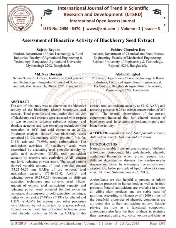
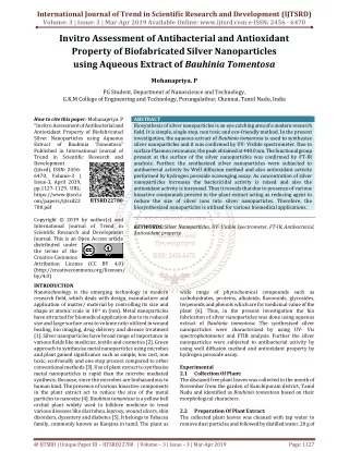
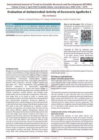
31 Int. J. Biomol. Biomed. groups were determined by One-way analysis of increase in SOD activity in negative control (Group 2) variance (ANOVA) with Dunnett post-hoc test using compared to the normal control (Group 1). Treatment Graph pad prism 5.0. Significant difference was with LJECB at 400 and 800 mg/kg caused a considered if p < 0.05. significant two-fold increase in SOD when compared Results with the normal or negative control groups. The normal Group 6 and 7 that received only 400 or 800 Effect of LJECB on serum reduced glutathione level mg/kg extract also showed a significant increase in in normal and wistar rats with indomethacin SOD activity when compared with the normal control. activity Likewise the positive control (Group 3) treated with The effects of LJECB on serum GSH is shown in Fig. Gecrol antacid, high antioxidant SOD activity was 1. There was a significant (p<0.05) increase in serum also observed. GSH level of the negative control (Group 2) when compared with the normal control (Group 1). However, Group 3 (positive control) treated with Gecrol antacid showed higher level of GSH compared to the negative control and normal control groups. Interestingly, treatment with LJECB at 800 mg/kg significantly lowered the GSH level to normal. Normal Group 6 and 7 that received 400 and 800 mg/kg extract showed a significant increase in GSH level when compared with the normal control that Fig. 1. Effect of LJECB on serum reduced glutathione received only the distilled water. The increase in GSH level in normal and wistar rats with indomethacin levels after in vivo administration with LJECB extract activity suggests enhanced antioxidant defense mechanisms and its potential to combat oxidative stress and protect the cells against oxidative damage. Effect of LJECB on serum glutathione peroxidase activity in normal and wistar rats with indomethacin toxicity The effects of LJECB on serum GSH is shown in Fig. 2. There was a non-significant increase in serum GPx activity of the negative control (Group 2) when compared with the normal control (Group 1). The Fig. 2. Effect of LJECB on serum glutathione normal Group 7 that received 800 mg/kg extract peroxidase level in normal and Wistar rats with showed a non-significant increase in GPx activity indomethacin activity when compared with the normal control. Following LJECB treatment, there was a non-significant rise in GPx activity, indicating that the plant extract may have a minor effect on the antioxidant defence system in female Wistar rats. Effect of LJECB on serum superoxide dismutase (sod) activity in normal and wistar rats with indomethacin toxicity Fig. 3. Effect of LJECB on SOD activity in normal The effects of LJECB on serum SOD is shown in Fig. and Wistar rats with Indomethacin toxicity 3. Indomethacin administration induced a significant Anyim et al.
32 Int. J. Biomol. Biomed. Effect of LJECB on serum catalase activity in normal and wistar rats with indomethacin toxicity The effect of LJECB on serum CAT is shown in Fig. 4. Indomethacin administration induced a non- significant increase in catalase activity in the negative control group compared to the normal control. Treatment with the LJECB at 800 mg/kg caused a significant increase in the catalase activity compared with the normal control group. Fig. 4. Effect of LJECB on serum catalase activity in Effect of LJECB on serum malondialdehyde (mda) in normal and Wistar rats with indomethacin toxicity normal and wistar rats with indomethacin toxicity MDA, a product of lipid peroxidation, is used as a biomarker of oxidative stress. The effect of LJECB on MDA concentration is shown in Fig. 5. There was a significant decrease in MDA level in the negative control group when compared with the normal control (Group 1). However, treatment with the Gecrol antacid caused a significant increase in MDA level when compared with the normal or negative Fig. 5. Effect of LJECB on Serum MDA in normal control group. When treated with the LJECB, the and Wistar rats with indomethacin toxicity MDA level was restored to normal as in the normal control group. The normal groups in 6 and 7 however, showed a significant increase in MDA when compared with the normal or negative control groups. Suggesting that consumption of LJECB without underlying disease condition may trigger undue lipid peroxidation. Effect of LJECB on total protein in normal and wistar rats with indomethacin toxicity The effects of LJECB on protein concentration in the Fig. 6. Effect of LJECB on total protein serum and gastric content is shown in Fig. 6. concentration in normal and Wistar rats with Indomethacin administration induced a significant indomethacin toxicity increase in serum protein concentration in negative control (Group 2) compared to the normal control (Group 1). Treatment with gecrol (Group 3) caused a further spike in serum total protein concentration. However, administration of LJECB at 800 mg/kg caused a significant reduction in the serum total protein concentration. Administration of 400 or 800 mg/kg of LJECB to the animals in Group 6 and 7 produced no significant effect when compared with the normal control. The total protein concentration Fig. 7. Effect of LJECB on nitric oxide (NO) was found to be significantly higher in serum when concentration in normal and Wistar rats with compared with the gastric juice. indomethacin toxicity. Anyim et al.
33 Int. J. Biomol. Biomed. Effect of LJECB on nitric oxide (NO) concentration in Studies have shown that, a common denominator for normal and Wistar rats with indomethacin toxicity pathogenesis of gastric ulcer is free radical The NO is a gastric defensive or protective factor perturbation (El-Ashmawy, 2016). The most against indomethacin-mediated ulcer. The effect of important and viable first line of defense in LJECB on NO concentration in normal and Wistar combating oxidative stress is through the use of rats with indomethacin toxicity is shown in Fig. 7. endogenous antioxidants such as superoxide There was a significant decrease in the NO level in the dismutase (SOD), catalase (CAT), and glutathione negative control (Group 2) compared to the normal peroxidase (GPx) (Ighodaro et al., 2018). Basically, control (Group 1). Conversely, the positive control these enzymatic antioxidants help in the conversion group (Group 3) treated with Gecrol antacid restored of peroxides and hydroperoxides to water and oxygen the NO level to the normal level as compared with the molecules which are subsequently removed from the normal control. Group 4 and 5 treated with 400 and cells without the risk of causing molecular damage 800 mg/kg of LJECB showed a significant increase in (Ighodaro et al., 2018). The nonenzymatic NO level compared to the normal control. As a antioxidants, including reduced glutathione (GSH) defensive factor, the increase in nitric oxide level also protect the cells against oxidative damage by suggests some protective effects on the gastric serving as electron donors to the radicals (Cadet et vascular system. The normal rats in Group 6 that was al., 2017). Some antioxidant mechanisms, including administered with 400 mg/kg of LJECB showed a elevated level of nitric oxide have been shown to significant increase in NO level. While administration increase the mucosal content of prostaglandin and of 800 mg/kg of LJECB to normal rats in Group 7 mucus in the gastric mucosa which suggests cyto- showed a nonsignificant difference in NO protective effects. Indomethacin may mediate gastric concentration when compared with the normal or ulceration by creating an imbalance between negative control. offensive factors (pepsin, gastric acid, ROS) and defensive factors (prostaglandins, bicarbonate ions, Discussion mucin, growth factors, and nitric oxide) (Serafim et This present study investigated the effects of lime al., 2023). juice extract of cashew bark (LJECB) on serum antioxidant profile in female Wistar rats with This present study observed a significant increase in indomethacin toxicity. NSAIDs including the activities of antioxidant proteins (GSH, CAT, indomethacin produce damage to the gastro- SOD, and GPX) in indomethacin-induced group intestinal mucosa in both humans and animals. This (negative control), suggesting possible accumulation explains why their use is associated with high risk of of ROS. The study however, observed a decrease in hemorrhage, perforation of mucosal lining in NO level in the negative control group with ulcerative conditions (Yuji and Toshikazu, 2006). The indomethacin toxicity. The decrease in NO level is molecular mechanism that allows NSAIDs like consistent with previous studies, showing that indomethacin to propagate gastro-intestinal toxicity indomethacin inhibits NO production by inhibiting is through the non-selective inhibition of the activation of endothelial nitric oxide synthase cyclooxygenase 1 and cyclooxygenase 2. As a result, (eNOS) (Arumugam et al., 2014). the COX-I is not able to produce sufficient prostaglandins required for housekeeping function However, groups treated with LJECB only showed a (Botting, 2007), including maintenance of the considerably higher level of NO, GSH, CAT, SOD, stomach endothelial walls. Thus, leading to the GPX. The increase in NO level following the pathogenesis of gastric ulcer and hemorrhage (Enas administration of LJECB suggests a possible et al., 2024). Ulceration may likely provoke rapid protective effect on the gastric vascular system. Some release of reactive oxygen species (ROS) which may studies reported increase in NO level of ulcerated rat lead to oxidative stress (Danisman et al., 2023). (Raish et al., 2021) which negates our study present Anyim et al.
34 Int. J. Biomol. Biomed. finding. Moreover, treatment with Gecrol antacid was activities to be significantly high in the negative associated with increase in NO level. The effects of control compared with the normal. Instead, a non- antacid on nitric oxide level may vary depending on significant difference was observed in the activities of the specific antacid used and the mechanism both enzymes; suggesting that the indomethacin involved. It is possible that the antacid treatment may treatment may have affected the CAT and GPx via have influenced nitric oxide mechanism through different mechanisms. effects on gastric acid secretion. Considering the level of GSH, it was observed that the indomethacin-stressed negative control had Furthermore, MDA is used as a biomarker of lipid significantly higher level of GSH than the normal peroxidation-mediated oxidative stress. In this study, control group. Reduced glutathione is an antioxidant the negative control group was found to have a which confers bio-protection on cells. It has effective significantly lower level of MDA than the normal protection against toxic effects of substances on control group. Suggesting that intake of indomethacin may be associated with maintenance of cellular tissues. It has been reported that roughly 90% of all membrane health. However, treatment with Gecrol glutathione pool are stored in human system in was associated with elevated MDA level; indicating reduced form (GSH) and mobilized during oxidative stress. The observed increase in the indomethacin- that intake of gecrol may be associated with stressed negative control could possibly be to combat peroxidative stress on the gastric mucosal wall. When the toxic effect of indomethacin. Treatment with 800 treated with the LJECB, the MDA level was restored mg/kg of LJECB showed a significant decrease in to normal as in the normal control group. GSH compared to the negative and normal control Administration of 400 or 800 mg/kg of LJECB to groups. normal rats in groups 6 and 7 was associated with a significant increase in MDA when compared with the Moreover, the total protein concentrations in both normal or negative control groups. Suggesting that serum and gastric juice were also compared. It was consumption of LJECB without an underlying observed that more proteins were present in the condition may trigger undue lipid peroxidation. serum than in the gastric juice. Indomethacin administration induced a significant increase in The observation that indomethacin-induced stress in serum protein concentration in negative control Wistar rats significantly elevated the SOD levels is in (Group 2) compared to the normal control (Group 1). agreement with previous studies (Danisman et al., Treatment with gecrol (Group 3) even caused a 2023). SOD has been shown to functionally transform further spike in serum total protein concentration. superoxide anion into hydrogen peroxides (H2O2) and However, administration of 800 mg/kg of LJECB was molecular oxygen (O2). An increase in SOD activity associated with a significant reduction in the serum suggests an increase in the intra-cellular total protein concentration. Administration of 400 or concentration of H2O2 resulting from superoxide 800 mg/kg of LJECB to the animals in Group 6 and 7 dismutation (Asma et al., 2014). produced no significant effect when compared with the normal control. The total protein concentration In contrast, indomethacin-induced stress caused a was found to be significantly higher in gastric content non-significant difference in catalase activity in the juice when compared with the serum. negative control group compared to the normal control. Treatment with the LJECB at 800 mg/kg was Indomethacin-induced stress was found to cause a associated with a significant increase in the CAT significant reduction in lipid peroxidation (MDA), activity. CAT is known to convert H2O2 to H2O; and NO levels; as well as an increase in the likewise, the GPx. considering the possible abundance antioxidant status. Administration of LJECB in of H2O2 from superoxide dismutase activity (Ray and female Wistar rats with indomethacin stress for 14 Husain, 20002). One expected the CAT and GPx Anyim et al.
35 Int. J. Biomol. Biomed. days, caused an increase in non-enzyme (NO and Asma B, Ranajoy C, Shankar M. 2014. Oxidative GSH) and enzymatic antioxidants (SOD, catalase) as stress: An essential factor in the pathogenesis of well as a non-significant decrease in GPX activity. gastrointestinal mucosal disease. Physiol. Rev. 94, While the plants’ extract elicited potent antioxidant 329–354. property, it was however, found to be associated with significant lipid peroxidative power in high doses. Ayala A, Munoz MF, Arguelles S. 2014. Lipid Therefore, prolong or frequent consumption of the peroxidation: production, metabolism, and signaling plants’ extract is strongly discouraged. mechanisms of malondialdehyde and 4-hydroxy-2- nonenal. Oxid. Med. Cell. Longev. 360438. Conclusion In conclusion, the LJECB elicited antioxidant Barbosa-Filho VM, Waczuk EP, Kamdem JP, property in the experimental rats. The increase in Abolaji AO, Lacerda SR, Da Costa JGM. 2014. nitric oxide levels suggested that there might be some Phytochemical constituents, antioxidant activity, protective effects on the gastric vascular system, while cytotoxicity and osmotic fragility effects of Caju the reductions in glutathione levels suggests that the (Anacardium microcarpum). Indus Crops Prod. 55, extract may also have some potential as a detoxifying 280–8. agent. However, the significant increase in malondialdehyde levels in ulcer-treated groups could Bicalho B, Rezende CM. 2001. Volatile be a cause of concern, as this may indicate increased compounds of cashew apple (Anacardium oxidative stress on the gastric endothelial cells. occidentale L.). Zeitschr Nat Sect C. 56,35–9. Therefore, it may be wise to exercise caution in using this extract, particularly at high doses or frequent Botting, R. 2007. Cyclooxygenases in Biology and dosage. Disease. In: Schmidt, R., Willis, W. (eds) Encyclopedia of Pain. Springer, Berlin, Heidelberg. References 978, 32. Adegbola MV, Godwin A, Ntwasa M, Ayeleso AO, Oyedepo TA. 2022. Potential Effect of Bryan NS, Grisham MB. 2007. Methods to detect Syzygium aromaticum (Cloves) Extract on Serum nitric oxide and its metabolites in biological Antioxidant Status and Lipid Profiles in Wistar Rats samples. Free. Radic. Biol. Med. 43, 645–657. with Artesunate Toxicity. Appl. Sci. 12, 8216. Cadet J, Davies KJA, Medeiros MHG, Di Aminjan HH, Abtahi SR, Hazrati, E, Mascio P, Wagner JR. 2016. Formation and repair Chamanara M, Jalili M, Paknejad B. 2019. of oxidatively generated damage in cellular DNA. Free Targeting of oxidative stress and inflammation Radic. Biol. Med. 107, 13–34. through ROS/NF-kappaB pathway in phosphine- induced hepatotoxicity mitigation. LifeSci. 232, Danisman B, Cicek B, Yildirim S, Bolat I, 116607. Kantar D, Golokhvast KS, Nikitovic D, Tsatsakis A, Taghizadehghalehjoughi A. 2023. Arumugam SN, Varghese J, Gautham TP, Carnosic Acid Ameliorates Indomethacin-Induced Visalakshi J, Molly J. 2014. Indomethacin inhibits Gastric Ulceration in Rats by Alleviating Oxidative activation of endothelial nitric oxide synthase in the Stress and Inflammation. Biomedicines 11(3),829. rat kidney: Possible role of this effect in the pathogenesis of indomethacin-induced renal damage. Ellman GL. 1959. Tissue sulfhydryl groups. Arch. Chemico-Biological Interactions 221, 77-87. Biochem. Biophys. 82, 70–77. Anyim et al.
36 Int. J. Biomol. Biomed. El-Ashmawy NE, Khedr E, El-Bahrawy HA, Liu S, Lou Y, Li Y, Zhang J, Li P, Yang B, Gu Q. Selim HM. 2016. Gastroprotective effect of garlic in 2022. Review of phytochemical and nutritional indomethacin induced gastric ulcer in rats. Nutrition characteristics and food applications of Citrus L. 32, 849–854. fruits. Frontiers in Nutrition 9,968604. Enas RA, Miada FA, Nada MA, Dalia HA, Azza Lowry OH, Rosebrough NJ, Farr AL, Randall MAZ, Faisal HA, Naseh AA. 2024. Apple extract RJ. 1951. Protein measurement with Folin-Phenol protects against indomethacin-induced gastric ulcers reagent. Journal of Biological Chemistry 193, 265- in rats by suppressing oxidative stress- The 275. implication of Nrf-2/HO-1 signaling pathway: In silico and in vivo studies, Journal of Functional Foods McCord JM, Fridovich I. 1969. Superoxide 112,105926. dismutase. An enzymic function for erythrocuprein Forman H J, Zhang H. 2021. Targeting oxidative (Hemocuprein). Journal of Biological Chemistry 244, 6049–6055. stress in disease: promise and limitations of antioxidant therapy. Nature Reviews Drug Discovery, Murphy MP, Bayir H, Belousov V. 20, 689–709. 2022. Guidelines for measuring reactive oxygen Fountoucidou P, Veskoukis AS, Kerasioti E, species and oxidative damage in cells and in vivo. Nat Docea AO, Taitzoglou IA, Liesivuori J. 2019. A Metab 4, 651–662. mixture of routinely encountered xenobiotics induces Özyürek M, Baki S, Güngör N, Çelik SE, Güçlü both redox adaptations and perturbations in blood and tissues of rats after a long-term low-dose K, Apak R. 2012. Determination of biothiols by a exposure regimen: The time and dose issue. Toxicol. novel on-line HPLC-DTNB assay with post-column Lett. 317, 24–44. detection. Anal. Chim. Acta 750, 173–181. Ighodaro OM, Akinloye OA. 2018. First line Pizzino G, Irrera N, Cucinotta M, Pallio G, defence antioxidants-superoxide dismutase (SOD), Mannino F, Arcoraci V, Squadrito F, Altavilla catalase (CAT) and glutathione peroxidase (GPX): D, Bitto A. 2017. Oxidative Stress: Harms and Their fundamental role in the entire antioxidant Benefits for Human Health. Oxid Med Cell Longev. defence grid. Alexandria Journal of Medicine 54, 2017,8416763. 287–293. Jaina S, Arora P, Popli H. 2020. A comprehensive Rahaman MM, Hossain R, Herrera-Bravo J, Islam MT, Atolani O, Adeyemi OS, Owolodun review on Citrus aurantifolia essential oil: its OA, Kambizi L, Daştan SD, Calina D, Sharifi- phytochemistry and pharmacological aspects. Rad J. 2023. Natural antioxidants from some fruits, Brazilian Journal of Natural Sciences 3, 354. seeds, foods, natural products, and associated health Jomova K, Raptova R, Alomar SY. 2023. benefits: An update. Food Sci Nutr. 11(4),1657-1670. Reactive oxygen species, toxicity, oxidative stress, and antioxidants: chronic diseases and aging. Arch Raish M, Shahid M, Bin Jardan YA, Ansari Toxicol 97, 2499–2574. MA, Alkharfy KM, Ahad A, Abdelrahman IA, Kumar P., Paramashivappa R, Vithayathil PJ, Ahmad A, Al-Jenoobi FI. 2021.Gastroprotective Effect of Sinapic Acid on Ethanol-Induced Gastric Subba Rao PV, Srinivasa Rao A. 2002.Process Ulcers in Rats: Involvement of Nrf2/HO-1 and NF-κB for isolation of cardanol from technical cashew Signaling and Antiapoptotic Role. Front Pharmacol. (Anacardium occidentale L.) nut shell liquid. J Agric 25(12),622815. Food Chem. 50,4705–8. Anyim et al.
37 Int. J. Biomol. Biomed. Ray G, Husain SA. 2002. Oxidants, antioxidants and carcinogenesis. Indian J Exp Biol. 40(11),1213-32. Sharifi-Rad M, Anil Kumar NV, Zucca P, Varoni EM, Dini L, Panzarini E, Rajkovic J, Sabiu S, Garuba T, Sunmonu T, Ajani E, Sulyman A, Nurain I, Balogun A. 2015. Indomethacin-induced gastric ulceration in rats: Protective roles of Spondias mombin and Ficus exasperata. Toxicol Rep. 8(2), 261-267. Tsouh Fokou PV, Azzini E, Peluso I, Prakash Mishra A, Nigam M, El Rayess Y, Beyrouthy ME, Polito L, Iriti M, Martins N, Martorell M, Docea AO, Setzer WN, Calina D, Cho WC, Sharifi-Rad J. 2020. Lifestyle, Oxidative Stress, Saleem M, Durani AI, Asari A, Ahmed M, Ahmad M, Yousaf N, Muddassar M. 2023 Investigation of antioxidant and antibacterial effects of citrus fruits peels extracts using different extracting agents: Phytochemical analysis with in silico studies. Heliyon. 9(4), e15433. and Antioxidants: Back and Forth in the Pathophysiology of Chronic Diseases. Front. Physiol. 11, 694. Sinha .K. 1972. Colorimetric assay of catalase. Anal. Bioche., 47, 389–394. Salehi B, Gültekin-Özgüven M, Kirkin C, Özçelik B, Morais-Braga Antioxidant, Antimicrobial and Anticancer Effects of Anacardium Plants: An Perspective. Frontiers in Endocrinology. Front. Endocrinol., 11. Statista 2023. Global leading lemon & lime MFB. 2020. producers 2022/23. https://www.statista.com/statistics/1045016/world- Ethnopharmacological lemons-and-limes-major-producers. Valko M, Leibfritz D, Moncol J, Cronin MT, Schmedes A, Holmer GA. 1989. A new thiobarbituric acid (TBA) method for determining free malondialdehyde (MDA) and hydroperoxides selectively as a measure of lipid peroxidation. Journal of Oil & Fat Industries 66, 813-817. Mazur M, Telser J. 2007. Free radicals and antioxidants in normal physiological functions and human disease. Int. J. Biochem. Cell Biol. 39(1), 44-84. Sen Li, Guowei Chen, Chao Zhang, Man Wu, Shuyan Wu, Qing Liu, 2014. Research progress of natural antioxidants in foods for the treatment of diseases, Food Science and Human Wellness 3(3-4), 110-116. Vona R, Pallotta L, Cappelletti M, Severi C, Matarrese P. 2021. The Impact of Oxidative Stress in Human Pathology: Focus on Gastrointestinal Disorders. Antioxidants 10, 201. Serafim C, Araruna ME, Júnior EA, Diniz M, Hiruma-Lima C, Batista L. 2020. A Review of the Role of Flavonoids in Peptic Ulcer (2010-2020). Molecules 25(22), 431. Yuji N, Toshikazu Y. 2006. Oxidative stress involvement and gene expression in indomethacin- induced gastropathy. Redox Report 11(6), 243-253. Anyim et al.