Download
1 / 6
0 likes | 16 Views
To evaluate the high-grade breast cancer morphological complexity on mammogram. We conducted a retrospective study using an open source data got from figshare repository. These anonymized data were collected and used for a study approved by the institutional review board. Cranio-Caudal and Medio-lateral mammograms and their tumor segmented images from 66 patients subdivided in two groups high histological grade (n=23) low-grade (low and intermediate, n=41). From breast cancer image segmentation, we extracted fractal dimension using Fraclac, plugin of ImageJ software based on box-counting

E N D
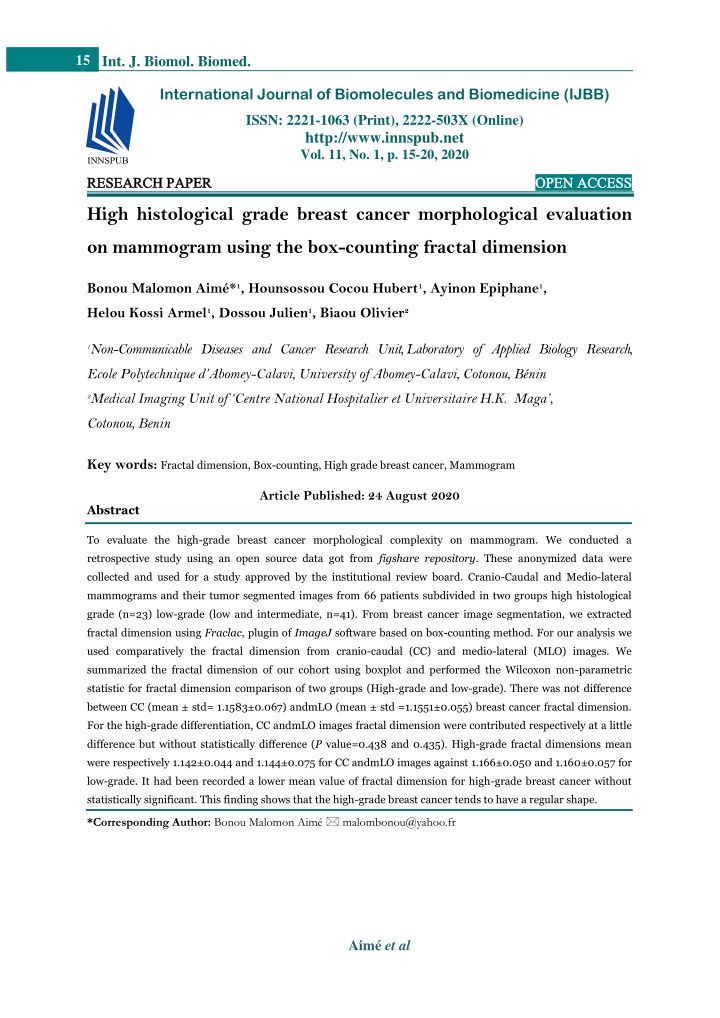
15 Int. J. Biomol. Biomed. International Journal of Biomolecules and Biomedicine (IJBB) ISSN: 2221-1063 (Print), 2222-503X (Online) http://www.innspub.net Vol. 11, No. 1, p. 15-20, 2020 RESEARCH PAPER RESEARCH PAPER OPEN ACCESS OPEN ACCESS High histological grade breast cancer morphological evaluation on mammogram using the box-counting fractal dimension Bonou Malomon Aimé*1, Hounsossou Cocou Hubert1, Ayinon Epiphane1, Helou Kossi Armel1, Dossou Julien1, Biaou Olivier2 1Non-Communicable Diseases and Cancer Research Unit, Laboratory of Applied Biology Research, Ecole Polytechnique d’Abomey-Calavi, University of Abomey-Calavi, Cotonou, Bénin 2Medical Imaging Unit of ‘Centre National Hospitalier et Universitaire H.K. Maga’, Cotonou, Benin Key words:Fractal dimension, Box-counting, High grade breast cancer, Mammogram Article Published: 24 August 2020 Abstract Toevaluate the high-grade breast cancer morphological complexity on mammogram. We conducted a retrospective study using an open source data got from figshare repository. These anonymized data were collected and used for a study approved by the institutional review board. Cranio-Caudal and Medio-lateral mammograms and their tumor segmented images from 66 patients subdivided in two groups high histological grade (n=23) low-grade (low and intermediate, n=41). From breast cancer image segmentation, we extracted fractal dimension using Fraclac, plugin of ImageJ software based on box-counting method. For our analysis we used comparatively the fractal dimension from cranio-caudal (CC) and medio-lateral (MLO) images. We summarized the fractal dimension of our cohort using boxplot and performed the Wilcoxon non-parametric statistic for fractal dimension comparison of two groups (High-grade and low-grade). There was not difference between CC (mean ± std= 1.1583±0.067) andmLO (mean ± std =1.1551±0.055) breast cancer fractal dimension. For the high-grade differentiation, CC andmLO images fractal dimension were contributed respectively at a little difference but without statistically difference (P value=0.438 and 0.435). High-grade fractal dimensions mean were respectively 1.142±0.044 and 1.144±0.075 for CC andmLO images against 1.166±0.050 and 1.160±0.057 for low-grade. It had been recorded a lower mean value of fractal dimension for high-grade breast cancer without statistically significant. This finding shows that the high-grade breast cancer tends to have a regular shape. *Corresponding Author: Bonou Malomon Aimé malombonou@yahoo.fr Aimé et al
16 Int. J. Biomol. Biomed. histological high-grade breast cancer tends to have a particular margin. Due to development the Computer Aid Diagnosis Introduction (CAD) based on mammography several reliable Breast cancer is the most common cancer in women quantitative features had been used to describe breast and a leading cause of cancer death worldwide (Bray cancer morphological characteristic. In this context, et al., 2018). Management of breast cancer relies on shape factors such as compactness, fractional the availability of robust clinical and pathological concavity, spiculation index, and a Fourier- prognostic and predictive factors to guide patient descriptor-based factor have been proposed for breast decision making and the selection of treatment. lesion classification (Rangayyan et al. 1997, 2000). Histological grade is one of important prognostic Latter fractal dimension had been used in the same factor. It is based on the degree of differentiation of purpose and it allowed to get a result better than with the tumor tissue and based on the evaluation of three previous features for the breast cancer differentiation morphological features: (a) degree of tubule or gland from benign lesion (Rangayyan and Nguyen 2007). formation, (b) nuclear pleomorphism, and (c) mitotic Fractal geometry is a powerful tool for describing and count. It is used to categorize breast cancer patients modeling natural objects. Most of these applications in three clinical groups grade I (low), grade II employ fractal dimension, a measure that captures (intermediate) and grade III (high) (Elston and Ellis the so-called complexity of the object, a fundamental 1991). High-grade breast cancer is recognized as more descriptor of analyzed objects represented in a digital aggressive cancer type and is the worst survival image. In this context, complexity expresses the level prognostic and need a specific treatment (WHO of detail detected at different scales. This measure is 2006; Rakha et al., 2008b, a). immediately related to physical characteristics, which are fundamental to the description and identification To date, the histological grading is one of popular of objects, even in our human vision system (texture method used to categorize breast cancer patients in analysis using fractal). In last decade, following therapeutic groups (low and high risk). Whereas, this success of CAD, several studies used medical image method has been described as subjective method with quantitative features in order to decrypt cancer sometimes inter-observer variability (Gilchrist et al., biology (Sanduleanu et al., 2018). Recently Fan et al. 1985; Theissig et al., 1990). and Huang et al. extracted quantitative features from In this context, some authors attempted to describe medical image to find those which are relevant to breast cancer histological grade (Huang et al., 2018; the high-grade breast cancer aspect on medical image Fan et al., 2019). In these previous studies, fractal in order to allow its a better identification for the dimension was not used, while it showed a better clinician. Regarding mammogram, Lamb et al. found potential for the differentiation of the breast tumors that classical appearance of a low or intermediate in according to their margin characteristic. Based on grade tumor is a speculated mass on mammography hypothesis that the high-grade breast cancer presents (Lamb et al., 2000). SHIN et al. 2011 had also a particular margin, we used in this study, the fractal attempted to describe it morphological aspect on dimension to evaluate its morphological complexity on mammogram and find the importance of this mammogram because mammography is one of the quantitative feature in its differentiation from other primary breast imaging modalities used in breast grades (low and intermediate). cancer diagnosis. They found that having Fairly slow Material and methods developing grade I tumors (low grade) and grade II tumors (intermediate grade) presents a stroma Patients data reaction resulting in imaging by spicules while high We conducted a retrospective study using an open grade with rapid evolution, do not develop a stroma source data got from figshare repository (Trevino reaction and has a round shape (Shin et al., 2011). 2018). These anonymized data were collected and The findings of both previous studies suggested that Aimé et al
17 Int. J. Biomol. Biomed. Fractal dimension determination used for a study approved by the institutional review board. It aimed to establish an association between digital Dicom Mammograms and tumor segmentation images were decompressed with the open source Dicom viewer mammography radiomic and breast cancer Oncotype software MicroDicom 2.7.9. Tumor segmented images DX and PAM50 recurrence scores. The study were rescaled between 0 and 1 grayscale with image englobes a total of 71 breast cancer cases with processing software ImageJ (Abràmoff et al., 2004; clinicopathologic informations (age, TNM grading, Schneider et al.,2012; ‘ImageJ, U. S. National ER, PR, and HER2 status), digital mammograms Institutes of Health, Bethesda, Maryland, USA, (cranio-caudal CC and medio-lateral obliquem LO), https://imagej.nih.gov/ij/, 1997-2018.’ 2018). We used microarray data and tumor segmentation on box-counting method to perform fractal dimension mammograms images. based on tumor segmented image. A digital mammography system (Selenia, Hologic, In box counting, data are gathered by laying boxes Bedford, MA), with an automatic intensity over a digital image as a series of grids of decreasing adjustment was used to acquire mammogram of 70 box size, then the number of boxes that fall on the microns per pixel and 12-bits grayscale for image (NC) and the size of each box (C) are recorded. codification. Manuel segmentation of tumors were C, the relative scale, can be considered as 1/box size, performed by an experienced breast radiologist because the image size is a constant. From this series (Tamez-Peña et al., 2018). Five (05) patients were of paired data, one infers the DB as the slope of the excluded because their histological grading status is log-log plot of C-1on the x -axis and NC on the y -axis missing. Amongst the sixty-six (66) patients of our (Fig. 1)(Karperien et al., 2008). In our study we used cohort, twenty-three (n=23) were high-grade, thirty- FracLac V.2.5 (Karperien 2015) a special ImageJ seven (n=37) were intermediate grade, and six (n=6) plugin for the box-counting fractal analysis. had low histological grading status with respective mean age of 50, 50.5 and 54 years. Fig 1. Fractal dimension extraction workflow. Statistical analysis groups (High grade and low grade). Statistical open For this step, we performed our analysis using source software R-3.4.2 had been used. All statistical comparatively the fractal dimension from cranio- tests were considered statistically significant if P caudal (CC) and medio-lateral (MLO) images. We value is less than 0.05. summarized the fractal dimension of our cohort using boxplot and performed the Wilcoxon non-parametric Results statistic for fractal dimension comparison of two Aimé et al
18 Int. J. Biomol. Biomed. Table 1shows that among twenty-three (23) high- grade breast cancer cases, eleven (11) had less than fifty (50) years old and twelve (12) were older than fifty (50) years. In low histological grade group we found twenty three (23) patients for under 50 years and twenty (20) for more than 50 years. In our cohort there is not a dependence between breast cancer histological grade and patients age. Table 1. Histological grade in function of patient age. Age<50 Yes 11 23 34 Histological grade High Low Total No 12 20 32 Total 23 43 66 Fig. 2. CC andmLO fractal dimension distribution. Fig. 2,3 and 4.shows respectively the fractal dimension distribution comparison between: •CC andmLO images of each patient; •high and low histological grade using CC image; •High and low histological grade usingmLO image. About the fractal dimension extracted from two projections (CC andmLO) of mammograms, there was not significant difference between the cohort mean (CC: mean ± std= 1.1583±0.067) (MLO: mean ±std =1.1551±0.055). For the high-grade differentiation, Fig. 3. histological high and low grade fractal there was no statistically significant difference between dimension using CC images. CC andmLO images fractal dimension (P value=0.438 and 0.435). High grade fractal dimension mean were respectively 1.142±0.044 and 1.144±0.075 for CC andmLO images against 1.166±0.050 and 1.160±0.057 for low grade breast cancer Table 2. Table 2. Fractal dimension of low- and high-grade breast cancer using CC andmLO images. Fractal Dimension Mean (±Std) High Grade 1.142(±0.044) 1.166(±0.050) 1.144(0.075) 1,160(±0.057) Low Grade P-value 0.438 0.435 CC Images MLO Images Fig. 4. histological high and low grade fractal dimension usingmLO images. Discussion Morphological analysis, in particular margin analysis of breast tumor on mammogram has contributed to a Aimé et al
19 Int. J. Biomol. Biomed. better characterization of its biology. It is well-known References that high-grade breast cancers show circumscribed Abràmoff DMD, Magalhães Dr PJ, Ram Dr SJ. margins because of their high cellularity and rich 2004. Image Processing with ImageJ 7. hyaluronic acid extracelluar matrix and inflammatory host reaction, whereas low-grade cancers show a Bray F, Ferlay J, Soerjomataram I, Siegel RL, spiculated margin because of their low cellularity, rich Torre LA, Jemal A. 2018. Global cancer statistics collagen matrix and desmoplastic host reaction 2018: GLOBOCAN estimates of incidence and (Stavros et al. 1995; Stavros 2004). Also Shin et al, mortality worldwide for 36 cancers in 185 countries. noticed that in their study high grade breast cancer CA: A Cancer Journal for Clinicians 68, 394-424. had the round shape resulting in its rapid evolution and low-grade presented irregular shape with spicule Elston CW, Ellis IO. 1991. Pathological prognostic because of its fairly development with stroma reaction factors in breast cancer. I. The value of histological grade (Shin et al. 2011). In our study, breast cancer with in breast cancer: experience from a large study with regular margin displayed fractal dimension low value long-term follow-up. Histopathology 19, 403-410. than breast cancer with irregular margin. Our result Fan M, Liu Z, Xie S, Xu M, Wang S, Gao X, Li were consistent to Rangayyan and Nguyen, who also L. 2019. Integration of dynamic contrast-enhanced reported that breast tumor with more irregular shape magnetic resonance imaging and T2-weighted had higher fractal dimension value (Rangayyan and imaging radiomic features by a canonical correlation Nguyen 2007). We observed that high-grade breast analysis-based feature fusion method to predict cancer fractal dimensions were lower than low histological grade in ductal breast carcinoma. Physics histological grade cancer, mainly using CC in Medicine & Biology 64, 215001. mammograms. This difference was not statistically significant, but it suggests that high-grade breast Gilchrist KW, Kalish L, Gould VE, Hirschl S, cancer presents more frequently regular margin than Imbriglia JE, Levy WM, Patchefsky AS, Penner low grade. DW, Pickren J, Roth JA, Schinella RA, Schwartz It is important to underline our study’s limitations. IS, Wheeler JE. 1985. Interobserver reproducibility of histopathological features in stage II breast cancer. Manual segmentation does not allow to find with Breast Cancer Research and Treatment 5, 3-10. more accuracy breast cancer margin on all mammograms mainly of young subjects who have Huang S, Franc BL, Harnish RJ, Liu G, Mitra denser breast. Mammography is planar medical D, Copeland TP, Arasu VA, Kornak J, Jones imaging modality leading the superposition of several EF, Behr SC, Hylton NM, Price ER, Esserman glandular structures with breast tumor. These two L, Seo Y. 2018. Exploration of PET and MRI realities contribute sometimes to the inaccessibility of radiomic features for decoding breast cancer the real breast cancer margin. The small size of our phenotypes and prognosis. NPJ Breast Cancer 4. cohort is also a limitation. Lamb PM, Perry NM, Vinnicombe SJ, Wells CA. In conclusion, in this study we used fractal dimension 2000. Correlation Between Ultrasound Characteristics, extracted from mammogram for differentiation of Mammographic Findings and Histological Grade in high histological grade breast cancer from low grade. Patients with Invasive Ductal Carcinoma of the Breast. We recorded a lower mean value of fractal dimension Clinical Radiology 55, 40-44. for high-grade breast cancer without statistically significant. This finding shows that the high-grade Rakha EA, El-Sayed ME, Lee AHS, Elston CW, breast cancer tends to have a regular shape. A future Grainge MJ, Hodi Z, Blamey RW, Ellis IO. large study will confirm our observations. 2008a. Prognostic significance of Nottingham Aimé et al
20 Int. J. Biomol. Biomed. histologic grade in invasive breast carcinoma. Journal Shin HJ, Kim HH, Huh MO, Kim MJ, Yi A, of Clinical Oncology: Official Journal of the American Kim H, Son BH, Ahn SH. 2011. Correlation Society of Clinical Oncology 26, 3153-3158. Rakha EA, El-Sayed ME, Powe DG, Green AR, between mammographic and sonographic findings and prognostic factors in patients with node-negative invasive breast cancer. The British Journal of Habashy H, Grainge MJ, Robertson JFR, Radiology 84, 19-30. Blamey R, Gee J, Nicholson RI, Lee AHS, Ellis Stavros AT. 2004. Breast Ultrasound. Lippincott IO. 2008b. Invasive lobular carcinoma of the breast: Williams & Wilkins. response to hormonal therapy and outcomes. European Stavros AT, Thickman D, Rapp CL, Dennis MA, Journal of Cancer (Oxford, England: 1990) 44. 73-83. Parker SH, Sisney GA. 1995. Solid breast nodules: use of sonography to distinguish between benign and Rangayyan RM, El-Faramawy NM, Desautels malignant lesions. Radiology 196, 123-134. JE, Alim OA. 1997. Measures of acutance and shape Tamez-Peña for classification of breast tumors. IEEE transactions J-G, Rodriguez-Rojas J-A, on medical imaging 16, 799-810. Gomez-Rueda H, Celaya-Padilla J-M, Rivera- Rangayyan RM, Mudigonda NR, Desautels JEL. Prieto R-A, Palacios-Corona R, Garza- 2000. Boundary modelling and shape analysis methods Montemayor M, Cardona-Huerta S, Treviño V. for classification of mammographic masses. Medical and 2018. Radiogenomics analysis identifies correlations Biological Engineering and Computing 38, 487-496. of digital mammography with clinical molecular Rangayyan RM, Nguyen TM. 2007. Fractal Analysis signatures in breast cancer. PLOS ONE 13, e0193871. Theissig F, Kunze KD, Haroske G, Meyer W. 1990. of Contours of Breast Masses in Mammograms. Journal of Digital Imaging 20, 223-237. Histological Grading of Breast Cancer: Interobserver, Sanduleanu S, Woodruff HC, Jong EEC, de, Reproducibility and Prognostic Significance. Pathology - Research and Practice 186, 732-736. Timmeren JE, van, Jochems A, Dubois L, Trevino V. 2018. Breast Cancer Images & Lambin P. 2018. Tracking tumor biology with radiomics: A systematic review utilizing a radiomics Segmentation - Correlation of Gene Expression quality score. Radiotherapy and Oncology 127, 349-360. Subtypes and Image Features. Schneider CA, Rasband WS, Eliceiri KW. 2012. WHO. 2006. Guidelines for management of breast NIH Image to ImageJ: 25 years of image analysis. cancer. World Health Organization, Regional Office Nature Methods 9, 671-675. for the Eastern Mediterranean, Cairo. Aimé et al