Download
1 / 10
0 likes | 8 Views
The extent of cryphonecrosis among the chestnut populations of three Imeretian (west Georgia) villages: Darka, Eto, and Chala has been evaluated. 23 strains of Cryphonectria parasitica (Murrill) Barr (syn. Endothia parasitica (Murrill) were isolated and identified from the bark of sick trees. The collection of strains of the plant pathogen fungus has been created. The strategy of the struggle against the chestnut blight, based on the application of antagonistic to C. parasitica microscopic fungi, has been elaborated. For this purpose 50 strains of different microscopic fungi were isolated

E N D
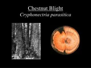
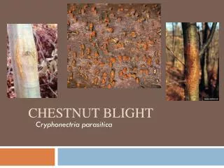
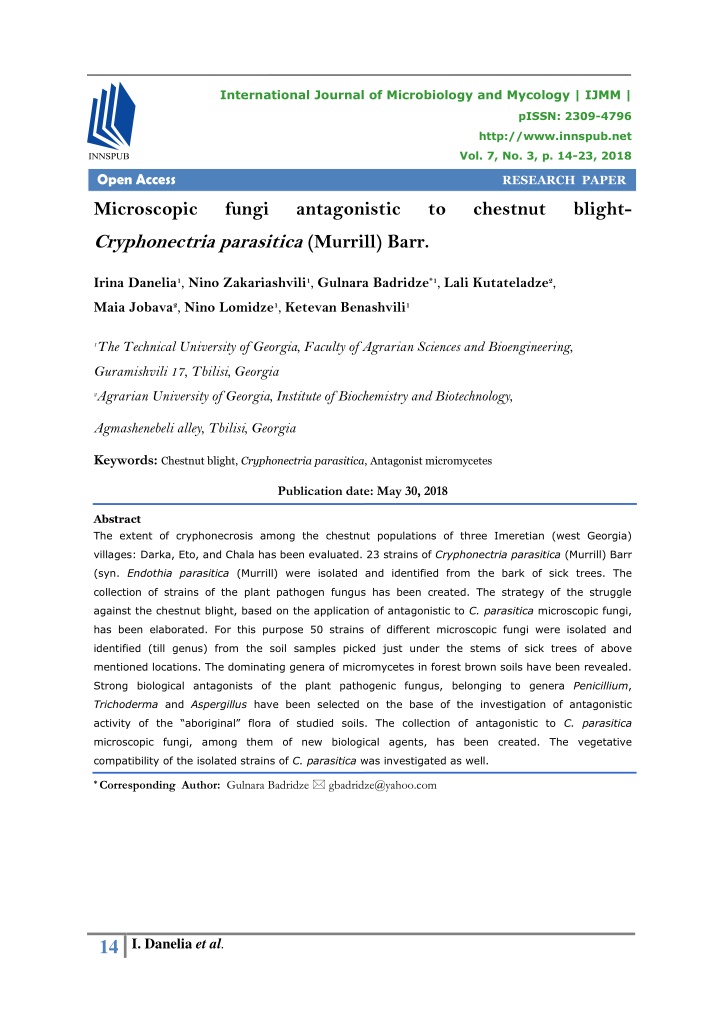
International Journal of Microbiology and Mycology | IJMM | pISSN: 2309-4796 http://www.innspub.net Vol. 7, No. 3, p. 14-23, 2018 Open Access RESEARCH PAPER Microscopic fungi antagonistic to chestnut blight- Cryphonectria parasitica (Murrill) Barr. Irina Danelia1, Nino Zakariashvili1, Gulnara Badridze*1, Lali Kutateladze2, Maia Jobava2, Nino Lomidze1, Ketevan Benashvili1 1The Technical University of Georgia, Faculty of Agrarian Sciences and Bioengineering, Guramishvili 17, Tbilisi, Georgia 2Agrarian University of Georgia, Institute of Biochemistry and Biotechnology, Agmashenebeli alley, Tbilisi, Georgia Keywords: Chestnut blight, Cryphonectria parasitica, Antagonist micromycetes Publication date: May 30, 2018 Abstract The extent of cryphonecrosis among the chestnut populations of three Imeretian (west Georgia) villages: Darka, Eto, and Chala has been evaluated. 23 strains of Cryphonectria parasitica (Murrill) Barr (syn. Endothia parasitica (Murrill) were isolated and identified from the bark of sick trees. The collection of strains of the plant pathogen fungus has been created. The strategy of the struggle against the chestnut blight, based on the application of antagonistic to C. parasitica microscopic fungi, has been elaborated. For this purpose 50 strains of different microscopic fungi were isolated and identified (till genus) from the soil samples picked just under the stems of sick trees of above mentioned locations. The dominating genera of micromycetes in forest brown soils have been revealed. Strong biological antagonists of the plant pathogenic fungus, belonging to genera Penicillium, Trichoderma and Aspergillus have been selectedon the base of the investigation of antagonistic activity of the “aboriginal” flora of studied soils. The collection of antagonistic to C. parasitica microscopic fungi, among them of new biological agents, has been created. The vegetative compatibility of the isolated strains of C. parasitica was investigated as well. * Corresponding Author: Gulnara Badridze gbadridze@yahoo.com 14 I. Danelia et al.
Introduction Combined application of antagonists and hypovirulent strains of C. parasitica appeared Wood of excellent quality and high nutritional especially effective against the disease (Akilli et value fruits rise the chestnut plant - Castanea al., 2011). Those countries which are unable to sativa Mill.in range of popular and economicaly control chestnut blight by hypovirulent strains, for significant trees in the world. (Heiniger and the purpose to localize the pathogen, apply the Rigling, 1994). Though, the plant is under the easy way of in situ struggle against the pathogen – danger of extinction all over the world. The reason soil compressing or mud packing method of it is so called “chestnut blight” disease caused (Anagnostakis, 1994; Groome et al., 2001). by the plant pathogenic fungus from ascomycetes The above mentioned botanical disaster of 20th Cryphonectria parasitica (Murrill) Barr (syn. Endothia parasitica (Murrill)) (Anagnostakis, 1994; century touched country of Geogia as well. Ten Rigling and Prospero, 2018). years ago 50% of Georgian chestnut forests were already dead (Prospero et al., 2013). There does It is more than half of a century the world not exist any effective fungicide against C. scientists try efforts to fight the chestnut blight parasitica, according to the information of but in vain. Healing of sick trees with Georgian Ministery of Environment Protection. hypovirulent strains is regarded to be the most Cutting of sick trees is the only way of effective measure against C. parasitica today cryphonecrosis localization here. Except the (Robin and Heiniger 2001; Puia et al., 2012). As joined scientific project of Swiss and Georgian it has established, hypovirulence is stipulated by investigators and attempts of the Turkish the existence of Cryphonectria hypovirus (CHV) scientists of integration of hypovirulent strains in in the cytoplasm of C. parasitica. The genome of Ajara chestnut forest, practically no active the virus is double chain RNA molecule (ds RNA). measures have been performed for saving of the The virus lacks of capsid, which suppresses its unique plant of Castanea sativa in Georgia, which active spreading in the environment. The virus is is placed in “Georgian red list” and “Red book” able “to enter” a new host organism only in case (Prospero et al., 2013). Thus, elaboration and of formation of the hypae anastomoses between testing of the strategy of biological control two stains of C. parasitica, or by means of the against cryphonecrosis in Georgian chestnut forests is very urgent task today. Accordingly, the host’s asexual spores. purpose of the presented work was to isolate the Though, wide scale integration of hypovirus under virulent strains of C. parasitica, spread in the natural conditions is not always possible chestnut populations of one of the regions of west because of the vegetative incompatibility of C. Georgia (Imereti), and to reveal the effective, parasitica’s different stains (Anagnostakis, 1977). antagonistic biological agents against them. The There exists another method of the biological strategy of the fight against chestnut blight, inspection of chestnut blight: isolation of spread in chestnut populations of west Georgian region Imereti, will be elaborated for the first antagonistic to C. parasitica microorganisms from time, and isolation of antagonistic to C. parasitica the natural sources and their application against microscopic fungi directly from one of the hot the pathogen. According to literary data among spots of cryphonecrosis of Georgia will be the antagonistic microorganisms of C. parasitica performed for the first time, as well as creation of are well known genera of microscopic fungi and the collection of antagonists against the bacteria (Trichoderma, Penicillium, Bacillus, pathogen. All this may be regarded as the Streptomyces) (Wilhelm et al., 1998; Groome et scientific novelty of the presented work. al., 2001; Akilli et al., 2011; Smith, 2013). 15 I. Danelia et al.
Collection of this type may be considered as the soil particles dispersion, microorganisms cells material base for management and localization of desorption and micro colonies separation (into a chestnut blight epidemic in this region. We hope particular composing cell) methods (Zvyagintsev that creation of the antinecrotic bio-preparation et al., 1980). As a result the homogenous and its in situ testing on the base of isolated suspensions were received. The suspensions were particular antagonist microscopic fungi or their cultivated by Waksman’s standard method of consortia will become possible in future. soils dilution (Waksman, 1916) and soils direct Materials and methods cultivation method (Warcup, 1950). Similar approach was applied while cultivating the Sampling suspension of bark samples. Following dilutions The experimental virulent strains of C. parasitica were prepared from the initial suspension: 1/10, were isolated from the sick chestnut trees of 1/100, 1/1000, and 1/10000. 0.1ml of the diluted Imereti region (west Georgia) chestnut forests, as suspensions was inoculated on Petri dishes with well as the cultures of microscopic fungi were the nutritional medium (g/l): 0.5l of wort 7oB, isolated from the soil samples picked just under the 0.5l of tap water, and 20 g of agar. The pH of the stems of sick trees of above mentioned location. medium was 5.5-6.0, sterilizing regimen – 45min, at 0.7atm. Pathogen was sampled by means of sterilized The initial inoculates were incubated in a lancers and knifes. Samples were taken in depth of 5mm from the infected bark of 8 sick trees, thermostat at 28o-30oC during 5days. The pure situated in the distance of 10m. For sampling the cultures were isolated after the primary antagonistic to C. parasitica microscopic fungi the microflora was received. Isolated from soil soil samples were taken in depth of 15cm in sick samples and purified, particular strains were chestnut forests (10 avareged samples) directly placed in test-tubes, on a sterilized, universal under the tree truks in the radius of 50cm, from agar nutrient medium and incubated in a five different places (Fomin, 2001). Samples were thermostat at 28o-30oC during 10days. Mature placed in sterilized containers and labled with cultures were stored in a fridge at 4oC, in test- indication of the location of sampling. tubes with the universal agar nutrient medium. Identification of microscopic fungi The initial inoculates and pure cultures From the beginning, till the identification of The initial Petri dishes cultures were obtained isolated fungi, the cultures were given the initial after the following procedures have been done: letters of the sampling location (E-Eto, D – first of all the suspensions of samples were Darka, Ch- Chala), as well as the number prepared. For this purpose one g of a particular (following numeration). On the first step of bark-sample was placed in a sterile 250ml conical identification the cultural-morphological vessel and was added with 99ml of tap water (the peculiarities of experimental fungi were studied initial suspension). To obtain relatively (speed of colonies growth, diameter, size, color, homogenous suspension of a particular etc.). Later the colonies were observed on Petri microorganism the diluted samples were placed dishes under the microscope, and preparations on shaker, at 30oC, 150rot/min. Almost the same for microscopy were prepared. procedures were performed to obtain the Part of the fungal colony, free of the agar of suspensions of soil samples, with the difference that 10g of each soil sample was added with nutrient medium, was cut out with the tip of 90ml of tap water (the initial suspension). Later sterilized loop and placed in water drop for the soil suspensions were treated by means of microscopic testing. 16 I. Danelia et al.
The spores spread on the surface of water drop. Determination of the vegetative compatibility of C. parasitica virulent strains Ethanol with ratio 1:1, or concentrated acetic acid The vegetative compatibility of the C. parasitica was added to water drop, as fungal spores and virulent strains (vc-types) was determined on Petri conidia do not adhere to water. The reprint- dishes as well. Preliminary sterilized agar nutrient preparation was prepared as well. For this purpose medium with potato and dextrose was inoculated 10mm in diameter colony was cut out from the agar side by side with particular virulent strains. Merging nutrient medium and placed on the preparation of different colonies, as well as formation of hyphae glass with the fungal colony upward. It was anastosomosis between virulent strains served as accurately and tightly covered with sterile cover indication to their compatibility. glass. Results and discussion Later the cover glass was placed on the preparation The visual evaluation of dissemination and extent of glass, which was previously dropped with water or cryphonecrosis in villages (Darka, Eto and Chala) of methylene blue. The prepared sample was Imereti region (west Georgia) was the initial step of investigated by the dry optical system. experiment. On the base of these observations the sampling individuals were selected. 23 strains of C. Screening of the microscopic fungi parasitica were isolated, purified and identified Particular culture of microscopic fungi, isolated from the experimental bark samples. Among from soils were inoculated in the same Petri dish, them 9 strains were from v. Eto chestnut forest, together with virulent strain of C. parasitica, on 10 – from v. Darka forest, and 4 strains – from v. the universal nutrient medium, for the purpose to Chala mixed deciduous forest (Fig. 1, 2). reveal antagonistic to chestnut blight microscopic Accordingly the collection of C. parsitica was fungi. The extent of inhibition of the growth of created. Guide of microscopic fungi was used for parasitic fungus indicated to the antagonistic the identification of pathogenic fungus activity of the neighbor, which was evaluated (F.M.Dugan, 2006; Malloch, et al. 1981). according to our 5 scale system. Fig. 1. The initial inoculates of the parasitic fungus C. parasitica, isolated from the infected chestnut trees. 17 I. Danelia et al.
Fig. 2. The pure, identified cultures of C. Parasitica. From literary data it is known that the plant morphotypically white, similar to hypovirulent pathogen fungus –C. parasitica may be infected strains, colonies were observed, which need by the dsRNA hypovirus (CHV-Cryphonectria further molecular investigation for identification. hypovirus), which deprives the pathogen of After the collection of C. parasitica strains was virulence and it becomes hypovirulent (Milgroom created, the vegetative compatibility between and Cortesi, 2004; MacDonald and Double, the strains was studied. Six vc types were 2004). The virulent strains of C. parasitica differ revealed, that indicates to high genetic diversity from the hypovirulent ones phenotipically as well. of the parasitic fungus of Imereti chestnut On agar medium with potato and dextrose (PDA) forests. The strains of C. parasitica of one they form yellow mycelia, with abundant spores. particular chestnut population appeared to be The pigmentation and sporulation of virus- incompatible with strains isolated from other infected C. parasitica is significantly low, chestnut populations as well. compared to virulent strains. On PDA they form white colonies. There has not been revealed any From the infected chestnut populations of Darka, clear hypovirulent strain of C. parasitica among Eto and Chala three incompatible groups of C. the isolated cultures. Though, in particular cases parasitica have been revealed (Fig. 3). Fig. 3. Three incompatible groups of C. parasitica from the infected chestnut populations of Darka, Eto and Chala. 18 I. Danelia et al.
Since the principle goal of our investigations was of reproductive organs and spores (Arx, 1970; to reveal the antagonistic to C. parasitica strains Kreisel and Fisher, 1969). For the identification of microscopic fungi under natural conditions, on on genus level were used guides after Pidoplichko the next step of our study 50 strains of and Milko (1971), Bilaiy and Koval (1988), antagonists were isolated and purified from Litvinov (1967), Malloch et al. (1981), and Fungal surrounding soils of chestnut trees. Among them Planet (2014). 20 strains were from Darkha soils, 19 – from Eto soils, and 11 strains – from Chala chestnut forest Identified microscopic fungi belonged to genera: soils (Fig.4). After the pure cultures were Mortierella, Verticillium, Fusarium, Trichoderma, received, their identification was important. At Trichothecium, Sporotrichum, Helminosporium, first the big taxonomic units (class, order) were Botritis, Cladosporium, Aspergillus, Penicillium, determined, according to structural peculiarities Mucor, Chaetomuim, Rhizopus. Fig. 4. The first inoculates of the microflora isolated from chestnut forest soils. Fungi Frequency in chestnut forest soils is demonstrated on Fig. 5. The dominating genera of microscopic fingi were revealed in soils of chestnut forests according to data on fungi frequency. Our experimental results are in accordance with literary data about dominance of genera Penicillium and Trichoderma in forest brown soils (Akilli et al., 2011; Groome et al., 2001; Smith, 2019). By frequency they are followed by genera Aspergillus and Fusarium. Quantitatively other genera of microscopic fungi Fig. 5. Microscopicfungi frequency in chestnut were presented in comparatively low amount. forest soils of Darka, Eto and Chala. 19 I. Danelia et al.
The identification of the microflora of pathogen (Akilli et al., 2011). It must be experimental soils has revealed a significant mentioned that new antagonistic to C. parasitica regularity: among those 50 strains of microscopic genus - Aspergillus was revealed in our fungi, which were isolated from tested soils C. experiments. No literary data exist on possibility parasitica was absent. This may be regarded as a of inhibition of the plant pathogen growth by this confirmation of the antagonistic activity of the fungus. Two species of Aspergillus isolated in our “aboriginal” microflora. The next step of experiments - A. Niger and A. Flavus totally experimental work aimed to test the antagonistic inhibited growth and development of C. parasitica effect of a particular strain of isolated strains (Fig. 6, b, c). During observations it was micromycetes towards C. parasitica. As it was established that the antagonistic effect of supposed, the representatives of genera micromycetes did not depend on a particular Penicillium and Trichoderma (Fig. 6, a) revealed location and was equal towards different strains high antagonistic effect towards the plant of C. parasitica (Table 1). Table 1. Screening of antagonistic to C. parasitica micromycetes C. parasitica strains Strains of soil micromycetes Aspergillus flavus-DY2 Aspergillus flavus-DY3 Aspergillus flavus-ET2 Aspergillus flavus-ET3 Aspergillus niger -W1 Aspergillus niger-DY1 Aspergillus niger-ET1 Botritis sp.-W2 Chaetomuim sp.-W3 10.Cladosporium sp.-DY4 11.Fusarium sp.-DY 17 12.Fusarium sp.-DY 18 13.Fusarium sp.-ET17 14.Fusarium sp.-ET18 15.Fusarium sp.-W10 16.Fusarium sp.-W11 17.Helminosporium sp.-E19 18.Mortierella sp.-ET5 19.Mucor sp.-DY5 20.Mucor sp.-DY6 21.Mucor sp.-ET 4 22.Penicillium sp.- ET7 23.Penicillium sp.-DY10 24.Penicillium sp.-DY11 25.Penicillium sp.-DY12 26.Penicillium sp.-DY7 27.Penicillium sp.-DY8 28.Penicillium sp.-DY9 29.Penicillium sp.-ET6 30.Penicillium sp.-ET8 31.Penicillium sp.-ET9 32.Penicillium sp.-W4 33.Penicillium sp.-W5 34.Rhizopus sp.-W6 35.Sporotrichumsp.-DY13 36.Trichoderma viride -W7 37.Trichoderma viride -W8 38.Trichoderma album -ET13 39.Trichoderma sp.-ET14 40.Trichoderma sp.-ET15 41.Trichoderma viride -DY14 C. parasitica ET- 1-9 C. parasitica W- 1-4 C. parasitica DY-1-10 No 1. 2. 3. 4. 5. 6. 7. 8. 9. ++++ +++++ +++++ +++++ +++ +++++ +++++ - - - ++ +++ +++ +++ - - - - - + + +++ +++++ ++ ++ ++ ++ +++++ - ++++ +++++ + +++++ - - +++++ +++++ - - - +++++ ++++ +++++ +++++ +++++ +++ ++++ +++++ - - - ++ +++ +++ +++ - - - - - + + +++ +++++ ++ ++ ++ ++ +++++ - ++++ +++++ + +++++ - - +++++ +++++ - - - +++++ ++++ +++++ +++++ ++++ +++ ++++ +++++ - - - ++ +++ +++ +++ - - - - - + + +++ +++++ ++ ++ ++ ++ +++++ + ++++ +++++ + +++++ - - +++++ +++++ - - - +++++ 20 I. Danelia et al.
42.Trichoderma viride -DY15 43.Trichoderma viride -ET11 44.Trichoderma viride -ET12 45.Trichoderma viride-DY16 46.Trichoderma viride-ET10 47.Trichoderma viride-W9 48.Trichothecium-ET16 49.Verticillium sp.-DY19 50.Verticillium sp-.DY20 +++++ ++ ++ ++ +++++ +++ - ++ ++ +++++ ++ ++ + +++++ +++ - ++ ++ +++++ ++ ++ ++ +++++ +++ - ++ ++ Collection of the antagonistic to C. parasitica microscopic fungi, created on the base of screening, may be concerned as the material base for management and localization of the chestnut blight epidemy. Creation of the antinecrotic preparation on the base of a particular antagonistic strain or their consortium and its in situ examination in future seems possible. Summarizing, the following significant results may be distinguished: 1. 23 strains of Cryphonectria parsitica (Murrill) Barrwere isolated, purified and identified from the cryphonecrosis-sick chestnut populations of three Imeretian (west Georgia) villages: Darka – 10 strains , Eto -9 strains, and Chala – 4 strains. The collection of the parasitic fungus has been created and the vegetative compatibility of the strains was investigated. 2. Six vegetatively incompatible (vc) types of C. parasitica were revealed. 3. 50 strains of microscopic fungi were isolated and purified from soils of infected chestnut forests for the purpose to reveal biological antagonists of C. parasitica. Among them 20 strains were from Darka soils, 19 – from Eto soils, and 11 strains- from Chala chestnut forest soils. Collection of mycromycetes was created. 4. The identified mycromycetes belonged to following genera: Mortierella, Verticillium, Fusarium, Trichoderma, Trichothecium, Sporotrichum, Helminosporium, Botritis, Fig. 6, a. representatives of genera Penicillium and Cladosporium, Aspergillus, Penicillium, Mucor, Trichoderma revealed high antagonistic effect Rhizopus and Chaetomuim. towards the plant pathogen. b, c. Two species of 5. Frequency of mycroscopic fungi in soils of Aspergillus - A. Niger and A. Flavus totally inhibited growth and development of C. parasitica strains. infected chestnut forests cleared the dominating genera of the first (Penicillium and Trichoderma) 21 I. Danelia et al.
and second order (Aspergillus and Fusarium). Dugan FM. 2006. The Identification of Fungi. An Other genera were presented in small amount. Illustrated Introduction with Keys, Glossary and 6. Investigation of the antagonistic effect of soil Guide to Literature. APS Press, St. Paul, MN. USA. 176. “aboriginal” microflora has revealed strong Fomin GS, Fomin AG. 2001.Soil.Monitoring on biological antagonists of the plant pathogen among particular strains of Penicillium and Quality and Ecologic Safety in Accordance with Trichoderma. International Standards. Moscow. VNII standard 7. New, antagonistic to C. parasitica genus – (in Russian). Groome PC, Tattar TA, Mount MS. 2001. Aspergillus has been revealed. Two species of this genus A. niger and A. flavus completely inhibited Bacteria found on American chestnut bark and growth and development of the virulent strains of their potential in biocontrol of chestnut the pathogen. blight. Arboricultural Journal 25(3), 221-234. Heiniger U, Rigling D. 1994. Biological control References Akilli S, Katircioğlu YZ, Maden S. 2009. of chestnut blight in Europe. Annual review of Vegetative compatibility types of Cryphonectria phytopathology 32(1), 581-599. parasitica, causal agent of chestnut blight, in the Kreisel H, Fisher G. 1969. Grundzuge eines Black Sea region of Turkey. Forest Pathology haturlichen systems der pilze. Jena. 39(6), 390-396. Litvinov A. 1967. Guide of microscopic soil fungi. Akilli S, Katircioğlu YZ, Maden S. 2011. Leningrad (in Russian). Biological control of chestnut canker caused by MacDonald WL, Double ML. 2004. Cryphonectria parasitica, by antagonistic organisms Hypovirulence: use and limitations as a chestnut and hypovirulent isolates. Turkish Journal of blight biological control. In: Restoration of Agriculture and Forestry 35(5), 515-523. American chestnut to forest lands. Proceedings of Anagnostakis SL. 1977. Vegetative incompatibility a Conference and Workshop 7-95. in Endothia parasitica. Experimental Mycology Malloch D. 1981. Moulds, their isolation, 1(4), 306-316. cultivation, and identification. University of Anagnostakis SL. 1994. Protecting chestnut Toronto Press. trees from blight. Northern Nut Growers Milgroom MG, Cortesi P. 2004. Biological control Association (USA). Arx, von JA. 1970. The genera of fungi of chestnut blight with hypovirulence: a critical analysis. Annu. Rev. Phytopathol 42, 311-338. Pidoplichko NM, Mylko AA. 1971.Atlas of sporulating in pure culture. Gramer Lehre, 1-288. Mucosal Fungi. Kiev, Naukova Dumka (in Russian). Bilaiy VI, Koval EZ. 1988.Aspergills. Kiev, Prospero S, Lutz A, Tavadze B, Supatashvili Naukova Doumka (in Russian). A, Rigling D. 2013. Discovery of a new gene Crous PW, Shivas RG, Quaedvlieg W, Van der pool and a high genetic diversity of the chestnut Bank M, Zhang Y, Summerell BA, Guarro J, blight fungus Cryphonectria parasitica in Wingfield MJ, Wood AR, Alfenas AC, Braun Caucasian Georgia. Infection, Genetics and U. 2014. Fungal Planet. Evolution 20, 131-139. 22 I. Danelia et al.
Puia CE, Grogorescu DA, Miclea RV. 2012. The Waksman SA, Curtis RE. 1916. The actinomyces Morphology and the Biological Control of of the soil. Soil Science 1(2), 99-134. Cryphonectria parasitica. Bulletin of the University Warcup JH. 1950. The soil-plate method for of Agricultural Sciences & Veterinary Medicine Cluj- isolation of fungi from soil. Nature 166, 117.-118. Napoca. Agriculture 69(1). Wilhelm E, Arthofer W, Schafleitner R, Krebs Rigling D, Prospero S. 2018. Cryphonectria B. 1998. Bacillus subtilis an endophyte of parasitica, the causal agent of chestnut blight: chestnut (Castanea sativa) as antagonist against Invasion history, population biology and disease chestnut blight (Cryphonectria parasitica). Plant control. Molecular plant pathology 19(1), 7-20. cell, tissue and organ culture 52(1-2), 105-108. Robin C, Heiniger U. 2001. Chestnut blight in Zviagintsev NDG. 1980. Methods of Soil Europe: diversity of Cryphonectria parasitica, Microbiology and Biochemistry. Moscow hypovirulence and biocontrol. Forest Snow and University, 12-14. (in Russian). Landscape Research 76(3), 361-367. Smith AR. 2013. Biological Control of Cryphonectria Parasitica with Streptomyces and an Analysis of Vegetative Compatibility Diversity of Cryphonectria Parasitica in Wisconsin, USA. Doctoral dissertation, University of Wisconsin-- La Crosse. 23 I. Danelia et al.