Download
1 / 7
100 likes | 595 Views
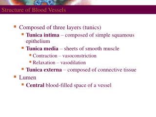
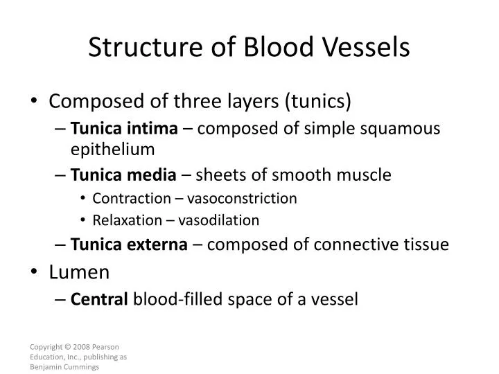
Structure of Blood Vessels. Composed of three layers (tunics) Tunica intima – composed of simple squamous epithelium Tunica media – sheets of smooth muscle Contraction – vasoconstriction Relaxation – vasodilation Tunica externa – composed of connective tissue Lumen

E N D
Structure of Blood Vessels • Composed of three layers (tunics) • Tunica intima – composed of simple squamous epithelium • Tunica media – sheets of smooth muscle • Contraction – vasoconstriction • Relaxation – vasodilation • Tunica externa – composed of connective tissue • Lumen • Central blood-filled space of a vessel
Structure of Arteries, Veins, and Capillaries Figure 19.1a
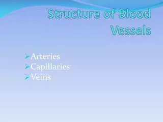
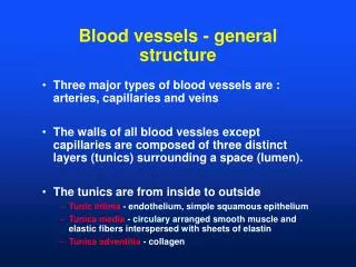
Types of Blood Vessels • Arteries – carry blood away from the heart • Capillaries – smallest blood vessels • The site of exchange of molecules between blood and tissue fluid • Veins – carry blood toward the heart
Types of Arteries • Elastic arteries – the largest arteries • Diameters range from 2.5 cm to 1 cm • Includes the aorta and its major branches • Sometimes called conducting arteries • High elastin content dampens surge of blood pressure Figure 19.2a
Types of Arteries • Muscular (distributing) arteries • Lie distal to elastic arteries • Diameters range from 1 cm to 0.3 mm • Includes most named arteries • Tunica media is thick • Unique features • Internal and external elastic laminae Figure 19.2b
Capillaries • Smallest blood vessels • Diameter from 8–10 µm • Red blood cells pass through single file • Site-specific functions of capillaries • Lungs – oxygen enters blood, carbon dioxide leaves • Small intestines – receive digested nutrients • Endocrine glands – pick up hormones • Kidneys – removal of nitrogenous wastes
RBCs in a Capillary Figure 19.3