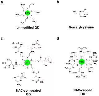
Download
1 / 21
210 likes | 395 Views
Imaging and spectroscopic performance studies of pixellated CdTe Timepix detector. Dima Maneuski. Vytautas Astromskas , Erik Fröjdh , Christer Fröjdh , Eva Gimenez -Navarro, Julien Marchal , Val O'Shea , Graeme Stewart, Nicola Tartoni , Heribert Wilhelm,

E N D
Imaging and spectroscopic performance studies of pixellated CdTe Timepix detector Dima Maneuski VytautasAstromskas, Erik Fröjdh, ChristerFröjdh, Eva Gimenez-Navarro, JulienMarchal, Val O'Shea, Graeme Stewart, Nicola Tartoni, HeribertWilhelm, Kenneth Wraight, RasifModhZain.
Table of contents • Presentation plan • Introduction • CdTe Timepix detector • Energy calibration • Diamond Light Source experiment • Laboratory X-ray tube experiment • Results • Energy resolution • Imaging performance • Charge sharing • Defects studies • Conclusions Dima Maneuski, PSD2011
CdTe sensor • Basic CdTe sensor properties • CdTe from ACRORAD • Bump-bonded to Timepix by FMF Freiburg • 1 mm thickness • 55 and 110 mm pixel pitch • Ohmic contacts (Pt) µeτe= 1.95 10-3cm²/Vs µhτh= 0.75 10-4cm²/Vs Dima Maneuski, PSD2011
Timepix detector • Timepix detector basic properties • 15 x 6 x 2 cm assembly size • Detector 14x14 mm, 256x256 pixels • 55 mm pixel pitch • ~550 transistors/pixel • 13.5 mW static power consumption • Up to 100 MHz ToT Clock • USB2.0 FitPix readout (~80 fps) • Operation modes • Counting • Time-over-threshold • Time-of-arrival Dima Maneuski, PSD2011
Signal clustering • Charge sharing • Fluorescence (Cd K-absorption edge – 26.7 keV, TeK-absorption edge – 31.8 keV) • Clustering is essential (software) • Clusters are between 55 and 2500 mm for 4 – 1000 keV Dima Maneuski, PSD2011
Energy calibration • Energy calibration procedure • 48 MHz Timepix clock • Single clusters identified • Non-linear function fitted • For energies > 100 keV • All clusters for calibration work better • Linear part of calibration only is needed For example Dima Maneuski, PSD2011
Diamond Light Source I15 • Extreme conditions beam line I15 • 48 hours allocated February 2011 • 20-80 keV • Beam size @ 40keV collimated by double slits to 20 mm • Energy resolution DE/E = 1x10⁻³ • Energies 25, 29, 33, 40 and 77 keV Dima Maneuski, PSD2011
Laboratory X-ray tube setup • Experimental setup • 55 and 110 mm detectors • Tungsten X-ray tube • Up to 50 keV • Up to 50 mA current • Various fluorescence metals (Ti, Ni, Cu, Zr, Ag, In, Sn) • Variable X-ray source (Rb, Mo, Ag, Ba, Tb, Am241) • Also Co57, Na22, Cs137, Co60 • PbNr slit for imaging X-rays • Default detector settings • -300V bias voltage • 48 MHz Timepix clock Dima Maneuski, PSD2011
55 mm pixel sources spectra Cs137 (662 keV) Mean 651 keV Sigma 55 keV DE/E = 8% Na22 (511 keV) Mean 494 keV Sigma 50 keV DE/E = 10% Dima Maneuski, PSD2011
110 mm pixel sources spectra Cs137 (662 keV) Mean 631 keV Sigma 34 keV DE/E = 5% Na22 (511 keV) Mean 480 keV Sigma 35 keV DE/E = 7% Dima Maneuski, PSD2011
110 mm pixel energy resolutions Diamond 25 keV 33 keV 77 keV Mean 80.2 keV Sigma 3.3 keV DE/E = 4% 29 keV 40 keV Dima Maneuski, PSD2011
Energy resolutions 55 & 110 mm pixel • Energy resolution for 110 mm pixel pitch is systematically better than for 55 mm pixel • @60 keV 7% vs. 13% • @662 keV 5% vs. 8% • Most likely due to additional pixel-2-pixel non-uniformities Dima Maneuski, PSD2011
Imaging performance (MTF’s) • Experiment • 60 keV X-ray tube • 55 mm pixel detector • Counting mode -300V -50V • Results • Optimal bias for imaging is > 400V • MTF varies 10-20% between regions in the sensoreven @ high biases Dima Maneuski, PSD2011
Imaging performance (MTF’s) Various X-ray tube energies • Experiment • Counting mode • Various energies @ -300V • Various thresholds (Noise 5 keV, E/2, 3/4E) • 55 mm vs. 110 mm pixel pitch • Results • ~15% difference between 20 keV and 60 keV @ 5.0 lp/mm • <10% difference between 5 and 15 keV threshold @ 20 keV @ 5.0 lp/mm • Most likely due to non-optimal CdTe bias voltage • MTF is better by > x2 for 55 um @ 4 lp/mm 55 mm vs. 110 mm MFT X-ray tube energy 20 keV Dima Maneuski, PSD2011
Charge sharing studies 25 keV • Experiment • Monochromatic X-ray beam • Pixel scan across the pixel • Time-over-Threshold Mode • Software energy thresholds (above E/2, below E/2) 40 keV Dima Maneuski, PSD2011
25 keV pixel scan Threshold above noise (>5 keV) • Energy-2-counts conversion • Superimposed count profiles from neighbouring pixels (x-1, x, x+1) • Threshold applied Threshold below E/2 (< 12.5 keV) Threshold above E/2 (>12.5 keV) Dima Maneuski, PSD2011
40 keV pixel scan Threshold above noise (>5 keV) • Energy-2-counts conversion • Superimposed count profiles from neighbouring pixels (x-1, x, x+1) • Threshold imposed Threshold below E/2 (< 20 keV) Threshold above E/2 (>20 keV) Dima Maneuski, PSD2011
25 keV vs. 40 keV • Energy 40 keV, threshold below E/2 • Charge sharing + fluorescence • Energy 25 keV, threshold below E/2 • Charge sharing only Dima Maneuski, PSD2011
Defect studies -500V -300V • Experiment • 55 mm detector • Counting mode • 60 keV X-ray tube • Variable bias voltage 14 mm -150V -50V • Results • High bias voltage suppresses visibility of defects • Defects “travel” over time • Defects result in non-uniform electrical field Dima Maneuski, PSD2011
Defect studies • +300V • +500V • Results • Different defects are visible • Defects “travel” and “pulse” over time • Defects result in non-uniform electrical field • Afterimage remains for sometime (bias switch on/off/reverse doesn’t help) • +150V • +50V 14 mm Dima Maneuski, PSD2011
Conclusions • Conclusions • 55 mm and 110 mm pixel CdTe Timepix detectors were compared for imaging and spectroscopic applications • X-ray tube and sources spectra and MTF’s • Diamond light source spectra, charge sharing • Analysis of CdTe defects • Positively/negatively charged defects • E-field distortions imaged • Future work • Per-pixel energy calibration -> better energy resolution • Optimal bias -> better imaging • Fancy correction algorithms • A lot of ideas for potential applications • Wakefield accelerator • Radioisotope production • ???? Dima Maneuski, PSD2011