Download
1 / 48
490 likes | 615 Views
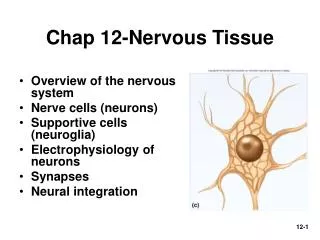
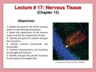
Chapter 12– Nervous Tissue. Ch. 12 Nervous Tissue– Study Guide. Critically read Chapter 12 pp. 442-461 before 12.5 Synapses. Comprehend Terminology (those in bold in the textbook) within the reading scope above

E N D
Ch. 12 Nervous Tissue– Study Guide Critically read Chapter 12 pp. 442-461 before 12.5 Synapses. Comprehend Terminology (those in bold in the textbook) within the reading scope above Study-- Figure questions, Think About It questions, and Before You Go On (section-ending) questions (within the reading scope above) . Before You Go On Questions. Do end-of-chapter questions— Testing Your Recall— 1-4, 7, 11-17 True or False– 1-4, 6, 8 10-2
§ Maintaining internal coordination by two body systems • Homeostasis? How? • Endocrine and nervous systems-- • Endocrine sys. –slower • chemical messengers (hormones) delivered to the bloodstream • Example-- insulin • Nervous sys. – quicker • chemical and electrical means • Example– in cold environment, vasoconstriction/shivering
§ Subdivisions of Nervous System Two major ANATOMICAL subdivisions: • Central nervous system (CNS) • brain and spinal cord enclosed in bony coverings • Peripheral nervous system (PNS)– all nervous tissue outside the CNS; made up of: • Nerves-- bundles of axons in connective tissue; emerge from the CNS; carry signals • Ganglia-- knotlike swellings in nerves Fig. @12.1
§ Functional divisions of PNS (SAME) • Sensory (Afferent) divisions (receptors to CNS)– carry signals to the CNS • somatic division—Ex. • visceral sensory division—Ex. • Motor (Efferent) division (CNS to effectors) • somatic motor division Effectors: skeletal muscles • visceral motor division (also called ANS) Effectors: glands and cardiac/smooth muscles • sympathetic division/parasympathetic division Fig. 12.2
PNS CNS Sensory division Brain Motor division Spinal cord Visceral motor division Somatic sensory division Somatic motor division Visceral sensory division Sympathetic division Parasympathetic division
§ Universal properties of neurons Nerves made up of nerve cells (neurons); neurons’ properties include: • Excitability • ability to respond to stimuli by producing action potential • Conductivity • produce traveling electrical signals • Secretion • Where? Why? • What is secreted?
§ Functional Classes of Neurons • Sensory (afferent) neurons • detect changes in body and external environment • Ex. • Interneurons (association neurons) • Confine ENTIRELY in CNS • 90% of our neurons are interneurons • process, store and retrieve information • Motor (efferent) neuron • send signals out to muscles and gland cells (effectors carry out body responses) • Ex. Fig. 12.3
Fig.12.3 Three Classes of Neurons Example--Detecting your own pulse at wrist
§ Neuron • Cell body = soma • Nucleus • Dendrites (1-many) • Function • Axon (single; nerve fiber) • Function Fig. 12.4 c-d-e
Neurofibrils Axon (d)
Schwann cell nucleus Axoplasm Axolemma Neurilemma Myelin sheath (c)
§ Variation in Neuron Structure– No. of processes from the soma: (Fig. 16.34) • Multipolar neuron • most common • Bipolar neuron • one dendrite/one axon • Unipolar neuron • Ex. sensory from skin to spinal cord directly • Anaxonic neuron • many dendrites/no axon • Ex. help in visual processes
§ Introduction • How many?! Neurons are outnumbered by neuroglia (1:50) in the nervous sys. • Functions- protect the neurons and help them function • Example– in the fetus, guide young migrating neurons to their destinations
§ 4 Types of Neuroglial Cells (CNS) • Astrocytes (star-shaped) • most abundant glial cells - form framework of CNS • contribute to blood-brain barrier and regulate composition of brain tissue fluid • Oligodendrocytes form myelin sheaths in CNS; distinguish these from Schwann cells • Ependymal cells (epithelial cells) line ventricles of the brain and central canal of the spinal cord; produce CSF • Microglia formed from monocytes; engulf invading microbes • in areas of infection, trauma or stroke Fig. 12.6
§ 2 Types of Neuroglial Cells (PNS) • Schwann cells-- myelinate fibers of PNS; assist in the regeneration of damaged fibers • Satellite cells – surround cell bodies in ganglia; regulate the chemical environment of the neurons
§ Myelin • Insulating layer around a nerve fiber; analogy– the rubber insulation on a wire • In CNS, • Each oligodendrocyte myelinate several fibers (Fig. 12.7a) • In PNS, • The ___________ cell wraps the nerve fiber • outermost coil is called neurilemma containing bulging body of the Schwann cell (nucleus and most of its cytoplasm) (Fig. 12.7b)
Myelin Sheath in CNS Figure 12.7b
Myelin Sheath in PNS Schwann cell Axon Myelin sheath Neurilemma
Myelin Sheath in PNS Node of Ranvier (gaps)-- between Schwann cells (also in CNS) Internodes– from one gap to the next
§ Unmyelinated nerve fibers • Locations? • CNS, PNS, both (circle one) • One Schwann cell harbors ______ small fibers • The Schwann cell– folds once around each fiber • Fig. 12.8
Unmyelinated nerve fibers Schwann cell Basal lamina
§ Myelination and Speed of Nerve Signal • Diameter of fiber and presence of myelin • large fibers have more surface area for signal conduction • Speeds • small, unmyelinated fibers = 0.5 - 2.0 m/sec • small, myelinated fibers = 3 - 15.0 m/sec • large, myelinated fibers = up to 120 m/sec • Functions • slow signals supply the stomach and dilate pupil • fast signals supply skeletal muscles and transport sensory signals for vision and balance
12.4A Electrophysiology of neurons KEY issues– How does a neuron generate an electrical signal? Cellular mechanisms for producing electrical potential and currents
§ Electrical Potentials & Currents • Electrical potential – a difference in the concentration of charged particles between one point and another • Electrical current– flow of charged particles from one point to another • Living cells have electrical potentials (are polarized) • resting membrane potential is -70 mV with a negative charge on the _______ of membrane; why? (next slide)
§ Resting Membrane Potential of all cells (RMP; -70 mV) Factors contribute to RMP: unequal distribution of electrolytes in ECF & ICF • Diffusion of ions down their conc. gradient • Selective permeability of the cell mem. • Cations and anions attract to each other Details-- • Membrane very permeable to K+ • Membrane much less permeable to Na+ • Cytoplasmic anions can not escape— EX. Proteins, phosphates etc. Fig. 12.11
Fig. 12.11--Ion basis of the resting membrane potential • Na+ more concentrated in the ECF • K+ more concentrated in the ICF Negative charge (-70 mV)
§ When a neuron is stimulated • Local potentials– changes in membrane potential when a neuron is stimulated • Causes– How? • Results–depolarization • Ionic bases-- • Na+ rushes into/out of (circle one) the cell • Na+ diffuses for short distance inside membrane producing a local potential Fig. 12.12 and X
For example– a chemical (pain signal; ligand) stimulates a neuron
Local potential-- Local, graded, and decremental Resting mem. Potential (-70 mV) Time Magnitude of stimulus Stimuli A B C D
§ Action potentials if stimulus is strong (Fig. 12.13) • Threshold reached • Depolarization (sodium channels open) • Repolarization (Sodium channels close and K+ gates fully open) • Hyperpolarization • Resting membrane potential restores 12-38
§ The refractory period of the action potential (AP) Period of resistance to stimulation for another AP • Absolute refractory period • as long as Na+ gates are open • no stimulus will trigger AP • Relative refractory period • as long as K+ gates are open • only especially strong stimulus will trigger new AP
12.4B--Conduction of a nerve signal in an unmyelinated fiber
Where are ion gates? • First action potential occurs at? • The next action potential occurs at? • Chain reactions continue until _____
§ Saltatory conduction (Fig. 12.17a-b) • Most (99%) of the voltage-regulated ion gates are at the _______________. • Slow but nondecremental • At the internodes– • nerve signals travel very ______ (diffusion of ions) and decremental. • Most of the axon is covered with myelin (internodes) • nerve signal is faster at 120m/sec (than unmyelinated ones (up to 2 m/sec)
Action potentials occurs only at the _________________ • It is called saltatory conduction meaning _____________.