Download

1 / 19
310 likes | 1.21k Views
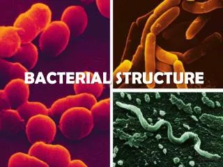
Bacterial Structure, Growth & Metabolism . Diagrammatic structure of a generalized bacterium. The cell envelope is composed of cell membrane – phospholipid bilayar ; acts a permeability barrier cell wall outer membrane (ONLY IN GRAM NEGATIVE BACTERIA) . . Bacterial cell wall.

E N D
Diagrammatic structure of a generalized bacterium • The cell envelope is composed of • cell membrane – phospholipidbilayar; acts a permeability barrier • cell wall • outer membrane (ONLY IN GRAM NEGATIVE BACTERIA).
Bacterial cell wall • Unique to bacteria • consists of the peptidoglycan layer & attached structures • Forms a strong mesh around the bacterial cells • Present in both Gram positive & Gram negative bacteria • Absent in Mycoplasma & Chlamydia • Important for • Maintaining the cell's shape • the rigid wall compensates for the flexibility of the phospholipid membrane • Countering the effects of osmotic pressure • Providing attachment sites for bacteriophages via the teichoic acids on the outer surface of the wall • Providing a rigid platform for surface appendages e.g flagella & pili
Peptidoglycan: Basic structure Muramic acid, D-amino acids & diamionpimelic acid are unique to bacteria • Glycan backbone: alternating monosaccharide subunits • N-acetyl muramic acid (NAM) & N-acetyl glucosamine (NAG) • Peptide side chains - made up of • D- and L- amino acids • Sometimes diaminopimelic acid • Peptide side chains cross link NAM subunits • Peptide bridges • cross-link the peptide side chains
Comparison of Gram positive & Gram negative cell envelopes Gram -ve: -thin peptidoglycan -Outer membrane lipopolysaccharide Braun lipoprotein Porin channels Gram +ve: -thick peptidoglycan -Lipotechoic & Teichoic acid
Principle of Gram stain Because of thin peptidoglycan cell wall, Gram negative bacteria lose the crystal violet after acetone decolourisation & take up the safranincounterstain Because of the thick peptidoglycan cell wall, Gram postive bacteria retain the Crytal violet dye after acetone decolourisation Gram positive bacteria stain bluish-purple Gram negative bacteria appear pinkish.
Bacterial surface structures Capsules Fimbriae (pili) Flagellae
Bacterial capsule • Found surrounding outside of cell envelope • not essential to cell viability • some bacteria may produce a capsule, others do not • It may be thick or thin • May be closely or loosely associated • Called slime layer when it is not well defined • Usually polysaccharide in nature • Functions of capsule:-important virulence factor • protects cell from desiccation and toxic materials • promote the concentration of nutrients at bacterial cell surface • play a role in adherence • protects bacteria from phagocytosis (killing) • capsular material is antigenic and helps in serological identification
Bacterial Surface Structures:The flagella • Long, filamentous appendages • Responsible for bacterial movement • Differ in their number and arrangement in the cell • single polar flagella –monotrichous • single flagella at two different poles –amphitrichous • two or more at one pole –lophotrichous • arising over the entire cell surface - peritrichous
Bacterial Surface Structures: Fimbriae (pili) • smaller appendages found on the surface of many bacteria • Shorter than flagella, thread-like • Composed of a protein • fimbrillin or pilin • Aid bacteria to adhere to one another – important virulence factor • Serve as attachment site for attachment to host cell surface • important for pathogenecity • Sex pili • involved in specific pair formation for exchange of genetic material during conjugation
Endospores • Dormant form of bacterial cell • The actively growing form is called vegetative • Produced by certain bacteria under adverse conditions (especially inadequate nutrients) • Spores are resistant to adverse conditions high temperatures & organic solvents • Survive under adverse conditions • Re-germinate when conditions are favourable • Commonly found in the genera Bacillus (e.g. Anthrax) and Clostridium (both Gram positive)
Endospore formation • Bacterial chromosomes replicates • Small amount of cytoplasm gathers with it • Cell membrane grows to seal off the developing spore • Once the walls of the spore are complete, spore is released
Endospores are difficult to eliminate • Most resistant living things known • Contain dipicolinic acid which helps stabilise their proteins and DNA • Makes it difficult to eliminate them from contaminated materials • Living spores have been recovered from Egyptian mummies • Can resist boiling for 2hours • Can survive in 70% ethanol for years • Highly resistant to radiation
Bacterial requirements for growth • Nutrients • All bacteria obtain energy by oxidising preformed organic molecules (carbohydrates, lipids and protein) from their environment • Metal Ions especially Iron • They secrete small molecules called siderophores • Siderophores bind iron which are then internalized into the bacterial cells via receptors • Ability to compete for iron is an impt virulence factor • Energy : ATP from metabolism of organic molecules • Optimal temperature • Many human bacteria are Mesophiles (grow at human body temp) • Some bacteria are pyschrophiles (survive at temp close to freezing) & some are thermophiles (survive at temp close to boiling) • Optimal pH : Many grow best at neutral pH ; Some can survive/grow in acid / alkali • Oxygen (or absence)
Environmental (O2) requirement for bacterial growth • Obligate Aerobes • Have an absolute or obligate requirement for oxygen e.g. Pseudomonas • Obligate Anaerobes • Cannot grow if any oxgen is present e.g. Bacteroides • Facultative anaerobes • Grow better if there is oxygen present but will also grow if oxygen is absent. • Facultative: means the organism is flexible in its oxygen requirement • E.g. Escherichia coli • Aerotolerant anaerobes • Also called obligate fermenters; They are indifferent to oxygen as they do not use it to transform energy. E.g. Streptococcus pyogenes; Lactobacillus • Microaerophilic • Require small amounts of oxygen for aerobic respiration (2-10%); Higher concentrations of oxygen are inhibitory E.g. Campylobacter & Helicobacter
Types of media In the laboratory we can grow bacteria on solid media (agar plates) or in liquid media (e.g. broth) Categories of media Complex media Differential media Selective media
TYPES OF MEDIA: DEFINITIONS • Complex media/ Enriched media : Contains a variety of ingredients for general bacterial growth e.g. Nutrient agar & Blood agar • Selective media: Substances e.g. antibiotics have been added suppress the growth of other organisms and only the desired organism will grow • Differential media: Contain substances that will change in a recognizable way for particular bacteria
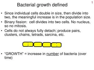
Kinetics of bacterial growth • Phases of growth • Lag phase ( cells adjusting to new environment • Exponential phase (cells growing exponentially) • Exponential growth via binary fission • Generation or doubling time (from 20 min to 24 h • Stationary phase ( rate of cell growth=rate of cell death) • Death phase (cells dying due to lack nutrition in media)