Download
1 / 86
970 likes | 1.38k Views
Bacterial Classification, Anatomy, Nutrition, Growth, Metabolism and Genetics. Classification Systems in the Prokaryotes. Macroscopic morphology C olony appearance & color Texture & size Microscopic morphology Cell shape, size Staining Physiological / biochemical characteristics

E N D
Bacterial Classification, Anatomy, Nutrition, Growth, Metabolism and Genetics
Classification Systems in the Prokaryotes • Macroscopic morphology • Colony appearance & color • Texture & size • Microscopic morphology • Cell shape, size • Staining • Physiological / biochemical characteristics • Enzymes • Chemical analysis • Chemical compound of cell wall • Serological analysis • Ag/ Ab binding • Genetic and molecular analysis • G + C base composition • Nucleic acid sequencing and rRNA analysis
G + C base composition • Low G+C Gram-Positive Bacteria • Clostridia • Mycoplasmas • High G+C Gram-Positive Bacteria • Corynebacterium • Mycobacterium
Bacterial Taxonomy Based on Bergey’s Manual • Bergey’s Manual of Determinative Bacteriology – five volume resource covering all known procaryotes • classification based on genetic information –phylogenetic • two domains: Archaea and Bacteria • five major subgroups with 25 different phyla
Major Taxonomic Groups of Bacteria • Vol 1A: Domain Archaea • primitive, adapted to extreme habitats and modes of nutrition • Vol 1B: Domain Bacteria • Vol 2-5: • 2 - Phylum Proteobacteria – Gram-negative cell walls • 3 - Phylum Firmicutes – mainly Gram-positive with low G + C content • 4 - Phylum Actinobacteria – Gram-positive with high G + C content • 5 – Loose assemblage of phyla – All gram negative
Species and Subspecies • Species • bacterial cells which share overall similar pattern of traits • Subspecies • Strain or variety • culture derived from a single parent that differs in structure or metabolism from other cultures of that species • E. coli O157:H7 • Type • subspecies that can show differences
Bacterial Shapes, Arrangements, and Sizes • Typically described by one of three basic shapes: • coccus • Spherical • bacillus • Rod • coccobacillus • vibrio • spirillum • Helical, twisted rod, • Spirochete
Bacterial Shapes, Arrangements, and Sizes • Arrangement of cells dependent on pattern of division and how cells remain attached after division: • cocci: • singles • diplococci • tetrads • chains • irregular clusters • cubical packets • bacilli: • chains • palisades
Bacilli Cocci
Appendages: Cell Extensions Flagella • 3 parts • filament • long, thin, helical structure composed of proteins • Hook • curved sheath • basal body • stack of rings firmly anchored in cell wall • rotates 360o • 1-2 or many distributed over entire cell
monotrichous single flagellum at one end lophotrichous small bunches arising from one end of cell amphitrichous flagella at both ends of cell peritrichous flagella dispersed over surface of cell, slowest Flagellar Arrangements
Fig. 4.4 Polar Rotates counterclockwise Cell swims forward in runs Reverse will stop it Peritrichous All flagella sweep towards one end Movement by flagella
aka Periplasmic Endoflagella Spirochetes enclosed between cell wall and cell membrane of spirochetes Internal Flagella Axial Filaments
Appendages for Attachment Fimbrae • fine hairlike bristles from the cell surface • function in adhesion to other cells and surfaces
rigid tubular structure made of pilin protein found only in Gram negative cells Functions joins bacterial cells for DNA transfer (conjugation) Adhesion to form biofilms and microcolonies Appendages for Mating Pili
The Cell Envelope • External covering outside the cytoplasm • Composed of few basic layers: • glycocalyx • cell wall • cell membrane • Maintains cell integrity
The Cell Membrane • fluid layer of phospholipid and protein • phospholipid molecules are arranged in a bilayer • Hydrophobic fatty acid chains in the phospholipids form a permeability barrier
Coating of molecules external to the cell wall Made of sugars and/or proteins functions attachment inhibits killing by white blood cells receptor The Bacterial Surface Coating Glycocalyx
2 types: slime layer - loosely organized and attached capsule - highly organized, tightly attached The Bacterial Surface Coating Glycocalyx
Cell Wall • Four Groups Based on Cell Wall Composition: • Gram positive cells • Gram negative cells • Bacteria without cell walls • Bacteria with chemically unique cell walls
macromolecule composed of a repeating framework of long glycan chains cross-linked by short peptide fragments provides strong, flexible support keep bacteria from bursting or collapsing because of changes in osmotic pressure Structure of the Cell Wall Peptidoglycan
Consists of a thick, homogenous sheath of peptidoglycan tightly bound acidic polysaccharides teichoic acid and lipoteichoic acid Periplasmic space cell membrane Gram Positive Cell Wall (1)
Consists of an outer membrane containing lipopolysaccharide (LPS) periplasmic space thin shell of peptidoglycan periplasmic space cell membrane Protective structure while providing some flexibility and sensitivity to lysis Gram Negative Cell Wall (2)
LPS endotoxin that may become toxic when released during infections may function as receptors and blocking immune response contains porin proteins in upper layer Regulates molecules entering and leaving cell Gram Negative Cell Wall
Important basis of bacterial classification and identification Practical aid in diagnosing infection and guiding drug treatment Differential stain Gram-negative lose crystal violet and stain red from safranin counterstain Gram-positive retain crystal violet and stain purple The Gram Stain
Atypical Cell Walls • Some bacterial groups lack typical cell wall structure • Mycobacterium and Nocardia • Gram-positive cell wall structure with lipid mycolic acid • pathogenicity • high degree of resistance to certain chemicals and dyes • basis for acid-fast stain • Some have no cell wall • Mycoplasma • cell wall is stabilized by sterols • pleomorphic
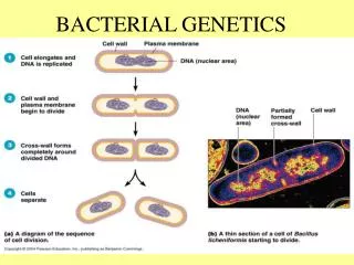
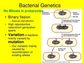
single, circular, double-stranded DNA molecule contains all the genetic information required by a cell DNA is tightly coiled around a protein dense area called the nucleoid central subcompartment in the cytoplasm where DNA aggregates Chromosome
Plasmids • small circular, double-stranded DNA • stable extrachromosomal DNA elements that carry nonessential genetic information • duplicated and passed on to offspring • replicate independently from the chromosome
Plasmids • may encode antibiotic resistance, tolerance to toxic metals, enzymes & toxins • used in genetic engineering • readily manipulated & transferred from cell to cell • F plasmids allow genetic material to be transferred from a donor cell to a recipient • R plasmids carry genes for resistance to antibiotics
intracellular storage bodies vary in size, number & content Examples: Glycogen poly-b-hydroxybutyrate gas vesicles for floating sulfur Storage Bodies Inclusions & Granules
resting, dormant cells produced by some G+ genera Clostridium, Bacillus & Sporosarcina resistance linked to high levels of calcium & certain acids longevity verges on immortality 25 to 250 million years pressurized steam at 120oC for 20-30 minutes will destroy Endospores
Endospores • have a 2-phase life cycle • vegetative cell • endospore • sporulation • formation of endospores • Germination • return to vegetative growth • withstand extremes in heat, drying, freezing, radiation & chemicals
stressed cell undergoes asymmetrical cell division creating small prespore and larger mother cell prespore contains: Cytoplasm DNA dipicolinic acid mother cell matures the prespore into an endospore then disintegrates environmental conditions are again favorable protective layers break down spore germinates into a vegetative cell Endospores
Obtaining Carbon • Heterotroph • organism that obtains carbon in an organic form made by other living organisms • proteins, carbohydrates, lipids and nucleic acids • Autotroph • an organism that uses CO2 (an inorganic gas) as its carbon source • not dependent on other living things
Growth Factors • organic compounds that cannot be synthesized by an organism & must be provided as a nutrient • essential amino acids, vitamins • Nutritional types • Chemo- • Chemical compounds • Photo- • light
Saprobes Parasites / pathogens Obligate Types of Heterotrophs
Nutritional Movement • Osmosis • Facilitated diffusion • Active transport • Endocytosis • Phagocytosis • Pinocytosis
digestion of complex nutrient material into simple, absorbable nutrients accomplished through the secretion of enzymes (exoenzymes) into the extracellular environment Extracellular Digestion
Environmental Influences on Microbial Growth • 1. temperature • 2. oxygen requirements • 3. pH • 4. Osmotic pressure • 5. UV light • 6. Barophiles
Minimum temperature lowest temperature that permits a microbe’s growth and metabolism Maximum temperature highest temperature that permits a microbe’s growth and metabolism Optimum temperature promotes the fastest rate of growth and metabolism 1. Temperatures
Psychrophiles optimum temperature 15oC capable of growth at 0 - 20oC Mesophiles optimum temperature 40oC Range 10o - 40oC (45) most human pathogens Thermophiles optimum temperature 60oC capable of growth at 40 - 70oC Hyperthermophiles Archaea that grow optimally above 80°C found in seafloor hot-water vents Temperature Adaptation Groups
Aerobe requires oxygen Obligate aerobe cannot grow without oxygen Anaerobe does not require oxygen Obligate anaerobe Facultative anaerobe and aerobe capable of growth in the absence OR presence of oxygen 2. Oxygen Requirements