Download
1 / 58
590 likes | 641 Views
ANATOMY OF PLACENTA PLACENTAL BARRIER. INTRODUCTION. Placenta is a remarkable organ Has a relative short life span, it undergoes rapid growth ,differentiation and maturation.

E N D
ANATOMY OF PLACENTA PLACENTAL BARRIER
INTRODUCTION • Placenta is a remarkable organ • Has a relative short life span, it undergoes rapid growth ,differentiation and maturation. • A unique fetal –maternal communication system which creates a hormonal environment that helps initially to maintain pregnancy and eventually initiates the events leading to parturition
The human placenta is: Discoid Hemochorial Deciduate Larynthine
Implantation is complete on 10 or 11th postovulatory day • On the 7th day ovum • Cytotrphoblast • Syncytiotrophoblast
Development of Placenta • Ovum • Morula • Blastocyst
Nitabuch’s layer • Placenta on 21 day of gestation –vascularised villous organ • The region of fibrinoid degeneration where the trophoblasts meet the decidua is calld nitabuchs layer. • This layer is absent in placenta accreta
Primary villi Secondary villi Tertiary villi
During this period there is some regression of the cytotrophoblastic elements in the chorionic plate and in the trophoblastic shell where cytotrophoblastic columns degenerate and largely replaced by fibrinoid material –Rohr’s layer
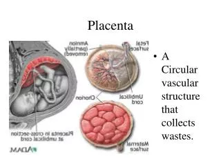
PLACENTA AT TERM: Placenta is a discoid organ 15 – 20cm in diameter 3cm Thick at center Weighs about 500gms
AT TERM MATERNAL SURFACE
Placental membrane • Total area-4 to 14 sq m • Similar to absorbtive area in adult git • In later part of pregnancy the membrane thickness reduces from 0.025mm to0.002mm • Is classified as haemochorial
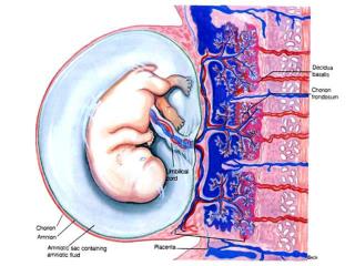
FETAL MEMBRANES 2 LAYERS: Outer chorion Inner amnion
Chorion Internally attached to amnion by loose areolar tissue Externally covered by trophoblastic layer and decidual cells of fused decidua capsularis and parietalis
Amnion Internal surface is smooth and shiny and in contact with liquor amnii Outer surface consists of a layer of connective tissue Amnion can be peeled off from the fetal surface of the placenta except at the insertion of the umbilical cord.
Development of membranes and formation of amniotic fluid • On the 8th and 9th postovulatory day • Endoderm • Ectoderm • Amniotic cavity • Primary yolk sac • Parietal extra embryonic mesenchyme • Visceral extra embryonic mesenchyme
Amniotic fluid • 12 weeks:50ml • 16 weeks:150ml • 38 weeks :900-1000ml • At term :1000-900ml • Clinical applications • Composition of amniotic fluid • functions
Placental circulation • Uteroplacental circulation • Circulation in the intervillous space • Feto-placental circulation
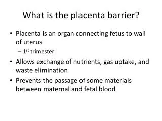
PLACENTAL BARRIER • Inspite of close proximity , there is no mixing of the maternal and fetal blood. • They are separated by placental membranes or barrier.
In early pregnancy it consists of: • Syncytiotrophoblast • cytotrophoblast • Basement membrane • Stromal tissue • Endothelium with fetal capillary wall • Its about 0.025 mm thick
Near term.. • Attenuation of syncytial layer • Sparse cytotrophoblast and distended capillaries fill the villus. • Vasculo- syncytial membrane: is the specialised zone of villi where the suncytiotrophoblast is thin and anuclear • These alphazones are for gas exchange. • Betazones of terminal villi are for hormone synthesis
BARRIER FUNCTION Fetal membrane is a protective barrier to the fetus against noxious agents circulating in the maternal blood. Antigen and antibody can traverse through the placental barrier The race of drug transfer is increased in late pregnancy
Maternal infections caused by : Virus Bacteria Protozoa , is transmitted to the fetus by crossing the placental barrier.
Placental functions • Simple diffusion • Facilitated diffusion • Active transfer • Endocytosis • Exocytosis • Respiratory functions • Excretory functions
Nutritive function • Enzymatic function • Barrier function • Immunological function • Hormones
Hormones produced by placenta • HCG • HUMAN PLACENTAL LACTOGEN • CHORIONIC ADRENOCORTICOTROPIN • RELAXIN • PARATHYROID HORMONE RELATED PROTEIN • GROWTH HORMONE RELATED VARIENT • HYPOTHALAMIC LIKE RELEASING HORMONE • GONADOTROPIN RELEASING HORMONE • CORTICOTROPIN RELEASING HORMONE
Clinical aspects of placenta • Multiple pregnancy
Placenta praevia • Normal sites of implantation of ovum • Upper uterine segment • Abnormal sites of implantation of ovum • Types of placenta praevia • First degree • Second degree • Third degree • Fourth degree
Bleeding following premature separation of normally situated placenta • Incidence: 0.49 to 1.8% • Types : concealed :20 to 35% revealed : 65 to 85%
Gestational trophoblastic disease • Proliferative abnormality of trophoblast associated with pregancy • Persistance GTD = GESTATIONAL TROPHOBLASTIC NEOPLASIA
Classification • Hyaditiform mole complete partial • Invasive moles • Placental site trophoblastic tumors • Choriocarcinoma • Non metastatic disease confirmed to uterus