Download
1 / 1
10 likes | 183 Views
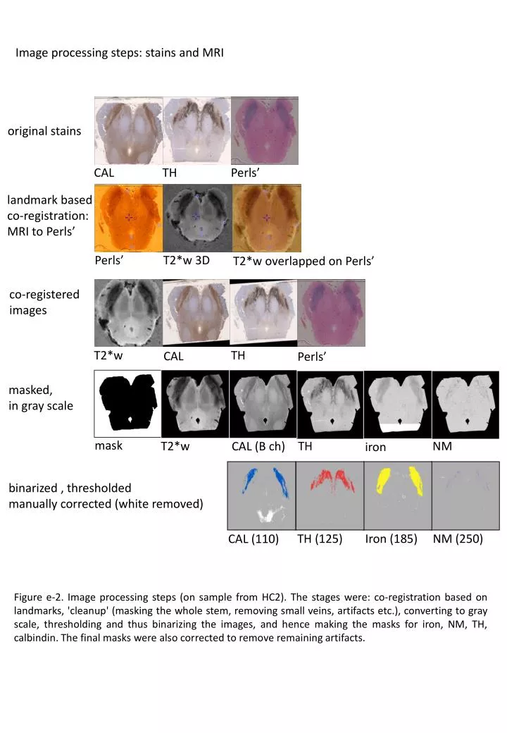
Image processing steps: stains and MRI. original stains. CAL. TH. Perls’. landmark based co-registration : MRI to Perls ’. Perls’. T2*w 3D. T2*w overlapped on Perls’. co-registered images. T2*w. TH. CAL. Perls’. masked, in gray scale. mask. T2*w. TH. NM. CAL (B ch). iron.

E N D
Image processing steps: stains and MRI original stains CAL TH Perls’ landmark based co-registration: MRI to Perls’ Perls’ T2*w 3D T2*w overlapped on Perls’ co-registered images T2*w TH CAL Perls’ masked, in gray scale mask T2*w TH NM CAL (B ch) iron binarized , thresholded manually corrected (white removed) Iron (185) TH (125) NM (250) CAL (110) Figure e-2. Image processing steps (on sample from HC2). The stages were: co-registration based on landmarks, 'cleanup' (masking the whole stem, removing small veins, artifacts etc.), converting to gray scale, thresholding and thus binarizing the images, and hence making the masks for iron, NM, TH, calbindin. The final masks were also corrected to remove remaining artifacts.