Download
1 / 58
690 likes | 1.52k Views
Restriction and Constriction. Nick Tehrani, MD. Restrictive Cardiomyopathy. EVOLVED As a bedside clinical diagnosis of constriction confirmed by right heart catheterization findings of constrictive physiology in patients who:. POROVED NOT TO HAVE ANY PERICARDIAL DISEASE.

E N D
Restriction and Constriction Nick Tehrani, MD
Restrictive Cardiomyopathy EVOLVED As a bedside clinical diagnosis of constriction confirmed by right heart catheterization findings of constrictive physiology in patients who: POROVED NOT TO HAVE ANY PERICARDIAL DISEASE
Utilitity of: Traditional Hemodynamic Criteria Traditional Hemodynamic Criteria: are of LIMITED Utility Hurrell DG, Nishimura RA, Higano ST, Appelton CP, Danielson GK, Holmes DR, Tajik AJ. Value of dynamic respiratory changes in left and right ventricular pressures for the diagnosis of constrictive pericarditis. Circ. 1996; 93:2007-2013
Restrictive Cardiomyopathy Represents an extreme form of Diastolic Dysfunction: Abnormal increase in Diastolic ventricular pressure impeding filling of the LV To NL EDV
Diastole: A historical view • Diastole as the passive interval between systolic events • Discovery of Frank-Starling mechanism: • LV-EDV, helps regulate the SV • Katz: • After MV opening, LV pressure continue to decline, despite LV volume incresase LV as an active suction pump in early diastole.
Diastolic Properties of the LV End of IVRT Active suction. ATP req’d To Re-uptake Ca++
Quantitative assessent of the 4 phases of Diastole: • IVRT>100 ms • Earliest diastolic ab-normality. • Impaired LV relax. • Filling pressures=NL • Dz. progression: • Decreased LV compliance, and • Increased filling pressure Late diastolic filling Diastasis Early diastolic filling IVRT
Quantitative assessment of the 4 phases of Diastole: • IVRT • Early filling: • DT < 130-180 ms • Interplay of Early and Late filling: • E:A ratios……. • If E at A> 20 cm/s => E/A unreliable • Tachycardia • PR prologgation • “A” wave duration……. Utilized in relation to PV “a” wave duration.
E:A ratios, as a Function of Age Impaired LV relaxation (IVRT ) Decreased LV Compliance Restrictive Pseudonormal
Pathophysiologic Similarity of:Restriction and Constriction Abnormal increase in ventricular pressure impeding filling of the LV To NL EDV Constriction Restriction Myocardial Disorder Pericardial Disorder
Anatomy • Lt. Atrium is not completely intrapericardial • All other cardiac chambers are completely intrapericardial • Pulmonary Veins are completely intrathoracic
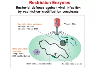
Effect of Inspiration • Normal Pericardium: • Inspiratory decrease in intrathoracic pressure is uniformly transmitted to the lungs, PVs, LA, LV, RA, and RV
Effect of Inspiration • Constrictive Pericarditis: • Thickened pericardium isolates the heart form transmission of intrathoracic pressure changes • Increased inspiratory capacitance of the Lungs PVs, and LA => PCWP decrease BUT • The decrease in intrathoracic pressure is not transmitted to the LV, RV, RA
Dissociation of Intrathoracic and Intracardiac Pressures First demonstrated to be present in constrictive pericarditis using Doppler techniques in 1989, by Hatle in her landmark study. Hatle LK, Appleton CP, Popp RL. Differentiation of constrictive pericarditis And restrictive cardiomyopathy by Doppler Echocardiography. Circ. 1989;79357-370
Dissociation of Intrathoracic and Intracardiac Pressures The inciting Physiologic Event. Hatle LK, et. al. Circ. 1989;79357-370
Ventricular Interdependence Hatle LK, et. al. Circ. 1989;79357-370 Ventricular Pressures Are DISCORDANT Insp Expir
Traditionalv.s.DynamicCatheterization Hemodynamics Why bother with Echo given The great utility of Dynamic Respiratory cath measurments? These measurments are only Possible using High-fidelity Micromanometer systems (not a common practice). Dissociation of Intrathoracic and Intracardiac Pressures
Effect of Inspiration:Constriction Inspir. Expir. Insp. Expir. PCWP PCWP Inspir. Expir. PCWP No proportionate decrease in LV diastolic pressure Decreased transmitral gradient => Transmitral flow RV SV LV SV
Pathophysiologic Differences Constriction Restriction Ab-Nl Myocardial compliance Myocardial compliance is NL No impedence to Diastolic EARLY FILLING Impedence to filling increases throughout the diastole Total cardiac volume is fixed by the pericardium Pericardium is compliant Septum is non-compliant Reduction of the proportion of LV filling with atrial contraction: => Atrial enlargement Atria are able to empty into the Ventricles, though at higher Press. Minimal Respiratory effect of RV on the LV Marked Respiratory effect of LV on the RV
SpecificEchocardiographic Criteria forConstriction/Restriction • Mitral E wave pattern • Pulmonary Vein pattern • Hepatic Vein pattern
Mitral E waveCriteria for Constriction • Decrease in of 25% in Mitral “E” velocity on inspiration.
In RESTRICTION: There is no respiratory variation of Mitral inflow
SpecificEchocardiographic Criteria forConstriction/Restriction • Mitral E wave pattern • Pulmonary Vein pattern
Normal PV Flow-TTE • PSV1- LA relaxation and pressure decrease. • PSV2- Interaction of RV-SV, w/ LA pressure and compliance. • PVa duration- Interplay of multiple factors Utilized in relation to Mitral “A” wave duration.
E:A ratios, as a function of Age Impaired LV relaxation (IVRT ) Decreased LV Compliance Pseudonormal Restrictive
Relation of Mitral “A” wave to Pulmonary Venous “a” wave duration • Normal Physiology • With LA contraction • Forward flowVolume and Duration Exceeds • Backward flow into the PV
Relation of Mitral “A” wave to PV “a” wave duration • RestrictivePhysiology: • PV-a Velocity > 35 cm/s OR • PV-a duration, 30 ms longer than Mitral “A” wave duration. 200 ms 121 ms
PVs are best assessed using TEE PV interrogation using TTE is often techniaclly limited.
Normal PV Flow-TEE Rt. Upper PV NO Variation from Inspiation to Expiration LV inflow
PV Dopplar Patterns in Restriction-TEE Lt. Upper PV PV flow is not respirophasic Systolic/Diastolic velocity is markedly down in both inspiration and expiration LV Inflow LV inflow Peak-E velocity is not respirophasic
PV Dopplar Patterns in Costriction-TEE Lt. Upper PV PV flowIS Respirophasic: • 25% variation of both the Systolic and Diastolic components Systolic/Diastolic • Ratio higher than for restriction (0.7 v.s. 0.4) LV Inflow LV inflow Peak-E: • 17% respiratory variation (v.s. none for restriction)
SpecificEchocardiographic Criteria forConstriction/Restriction • Mitral E wave pattern • Pulmonary Vein pattern • Hepatic Vein pattern
Respiratory Cycle :Hepatic Vein Flow IVC Inspiration Expiration
Hepatic Vein Dopplar: Normal Normal Systolic and diastolic forward flow S-vel. > D-vel. Diastolic flow reversal: Expir.>>Insp.
Hepatic Vein Dopplar: Constriction Constriction Diastolic flow reversal is augmented in expiration. DFRexp.>25% forward diastolic velocity
Hepatic Vein Dopplar: Restriction Restriction Forward flow primarily in Diastole. Inspiration increases both >systolic, and >Diastolic Flow reversals.
Nasser S Tehrani: These pts not respond as well to surgery Hepatic Vein Dopplar: Compilation Mixed physiology (restriction/constriction) Diastolic flow reversal during both Ispiration and expiration
Constriction Doppler Inspiration Expiration
Pitfalls and Caveats • Subgroup of patients with constriction who do not exhibit respiratory changes • COPD
Constriction: Non-respirophasic • Oh et. al. Circ. 1997;95:796-799 • 12 Pts. W/ confirmed constriction, but without the classic findings • Etiology of Non-respirophasicpattern • Mixed Restriction and Constriction • Marked increase in Preload Deduced post Stripping, as Sx Not improve Preload reduction to unmask the respiratory variation
Nasser S Tehrani:Wide STD.Deviation, But may be diagnostic for a ginven pt. Constriction: Non-respirophasic Supine Supine Sitting Insp. Expir. Insp. Expir. Sitting
Effect of changing loading conditions w/ VALSALVAin RESTRICTION E 20% A to a lesser degree
Pitfalls and Caveats • Subgroup of patients with constriction who do not exhibit respiratory changes • COPD
COPD v.s. Constriction • Individual Mitral flow velocity profiles are not restrictive as LV filling pressure is not increased. 100% change in E Velocity