Download
1 / 40
400 likes | 462 Views
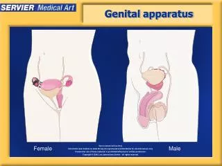
Explore the anatomy and functions of the female reproductive system, from the ovary, uterine tube, to the uterus. Learn about ovarian follicles, ovulation, corpus luteum, and the menstrual cycle. Dive into the details of endometrial layers, menstrual phases, and more.

E N D
summary • ovary: produce ovum & secret steroid hormones. • uterine tube: transport ovum+fertilization • uterus: where embryonic & fetal development occurs. • vagina • external genitalia
一、ovary • capsule:epithulium + albuginea. • parenchyma:cortex + medulla • cortex:ovarian follicles + CT. • medulla:LCT + elastic f + blood vessels
(一)ovarian follicle • Follicle = an oocyte + surrounding follicul cells. • 4 steps : primordial、primary、secondary and mature follicle 。
1. Primordial follicle • location: beneath the albuginea. • small & more. • primary oocyte + follicul C • primary oocyte : round , eosinophilic.
2. Primary follicle • primary oocyte. • Follicle C→proliferate,squamous→cuboidal or columnar,simple→stratified. • Zona pellucida: homogeneous.
3. Secondary follicle • Primary oocyte: size increase to the apex. • antrum: follicular fluid, 6~12 layers of granulosa cells.
Follicle C : granulosa C. • stratum granulosum:formed by several granulosa C , the wall. • Cumulus oophorus:primary oocyte + zona pellucida + corona radiata + granulosa C. protruding into the antrum
antrum Cumulus oophorus Granulosa cells
4. Mature follicle • secondary follicle→mature follicle • follicular fluid • the wall • move towards the surface of ovary. • primary oocyte →secondary oocyte
(二)ovulation • Mature follicle rupture,secondary oocyte + corona radiata + zona pellucida + follicular fluid, expelled from the follicle.
fate of secondary oocyte after ovulation • In 24h, degenerate and disappear if not fertilizing. • Or else,go on with the 2nd meiotic division,form haploid mature ovum.
(三)corpus luteum • Definition : after ovulation,the follicular wall, composed of the remaining granulosa & thecal cells , is thrown into deep folds as follicle collapse and is transformed into the corpus luteum. Show a yellow appearance in fresh preparations.
2. Functions of luteal cells • granular lutein C:progesterone & relaxin. • theca lutein C:estrogen
3. Classifications of corpus luteum • corpus luteum of menstruation : is formed in the absence of fertilization. • corpus luteum of pregnancy:is formed after fertilization and implantation.
二、uterine tube • infundibulum+ampulla+isthmus+uterine part • lumen is irregular • wall= mucosa+muscularis+serosa • mucosa= simple columnar Epi.+basal lamina • Epi.= secretory C + ciliated C • muscularis : inner circular, outer longitudinal SM
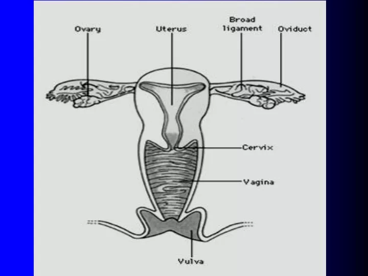
三、uterus • body+fundus+cervix • wall=endometrium+myometrium+perimetrium
1. endometrium • Function layer: superficial, undergo cyclical changes. • Basal layer: deeper, proliferate & repair function layer. • Epi=secretory C+ciliated C. • Basal lamina=CT+stroma C+ uterine gland . • Uterine gland: Epi. Invaginate to form. • Coiled artery: the branch of uterine artery.
2. menstrual cycle • cyclical changes of Function layer • exfoliate、bleed、repair & proliferate • occur every 28 days, called…。 • menstrual phase: first 1~4 days • proliferative phase: 5 ~ 14 • secretory phase: 15 ~ 28
1. proliferative phase • Also called follicular phase。 • Epithelial C+stroma C proliferate。 • Early : uterine gland sparse、thin & short. • later: endometrium become thick, uterine gland increase ,coiled artery go up and convolute. • Ovulation in overy,endometrium transform into the secretory phase.
2. secretory phase • Also called luteum phase. • Endometrium continue thickening. • Uterine gland highly convolute,swell,filled by secretion. • Stroma C hypertrophy. • Coiled artery increase,more convoluted. • If ovum is fertilized,endometrium keep thickening ,till into decidua,or else,into menstrual phase.
3. menstrual phase • If fertilized occurred,coiled A contract,endometrium ischemic lead to necrosis of function layer. • Brief periods of blood flow,blood flow into function layer. disruption of the surface epithelial C & rupture of the blood . Wash out patches of tissue separated from endometrium discharge through vaginalis.