Download
1 / 47
480 likes | 513 Views
Explore the latest advancements in diagnosing and treating neuroendocrine tumors, including carcinoids and endocrine pancreatic tumors. Learn about various diagnostic techniques like biochemistry, radiology, and somatostatin receptor scintigraphy. Stay updated on tumor biology, biomarkers, and treatment monitoring strategies.

E N D
Diagnosis and treatment of neuroendocrine tumors Dan Granberg
Carcinoids Bronchial Thymic Gastric Duodenal Small bowel Appendiceal Large bowel Rectal Endocrine pancreatic tumors Gastrinomas Insulinomas Glucagonomas VIPomas Somatostatinomas Non-functioning Mixed Neuroendocrine tumors
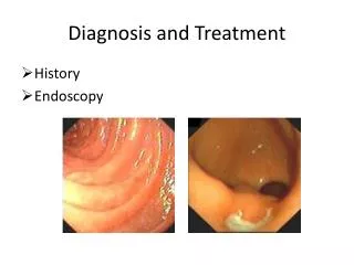
Diagnosis • Biochemistry • Radiology • CT • MRI • Ultrasonography • Endoscopic ultrasonography • Somatostatin receptor scintigraphy = octreoscan • Positron emission tomography = PET • Biopsy • Echocardiography • Endoscopy
Histopathology – Tumour biology • Neuroendocrine markers • Chromogranin A • Synaptophysin • Specific markers – gastrin, serotonin • Proliferation marker – Ki-67, PCNA • Adhesion molecules – CD44 • Angiogenic factors – VEGF, bFGF, TGFa • Tyrosine kinase receptors • Somatostatin receptors – SSTR 1-5
P-chromogranin A (P-chromogranin B) U-5’HIAA U-MeImAA P-ACTH U-cortisol S-gastrin S-PP (pancreatic polypeptide) P-glucagon P-VIP S-calcitonin S-insulin S-proinsulin S-C-peptide Secretin test Gastric pH 72-hour fasting Meal stimulation test Biochemistry
Biochemistry P-chromogranin A: • Most sensitive marker • Early detection of recurrence (Welin et al) • Treatment monitoring • Pitfalls • Impaired renal function • Treatment with proton pump inhibitors • Chronic atrophic gastritis • Inflammatory bowel disease • Decreased liver function • High spontaneous variation
Plasma chromogranin ASpontaneous variation Patients: Plasma chromogranin A measured on 2 consecutive days Granberg 1999
Plasma chromogranin ASpontaneous variation Results: Granberg 1999
Plasma chromogranin ASpontaneous variation Granberg 1999
Radiology • CT scan • native • i.v. contrast enhancement • late arterial phase = portal venous phase • venous phase • MRI • Ultrasonography • biopsy • Endoscopic ultrasonography • Intaoperative ultrasonography • Echocardiography • carcinoid heart disease
CT in neuroendolrine tumors 64 patients with GE-NETs Examined by CT, MRI and SRS In 40 pats (62,5%) liver metastases were found Maximum number of lesions detected for each patient (by SRS or CT or MRI) were added = Total number Relative sensitivity = number of lesions detected by method divided by total number of lesions. In a lesion-by-lesion analysis the sensitivities were: SRS 49% (204 mets) CT 79% (325 mets) MRI 95% (394 mets) Dromain 2005
Somatostatin receptor scintigraphy Neuroendocrine tumors: • Carcinoids • Midgut >90% • Bronchial 67% • Endocrine pancreatic tumors • Gastrinomas >90% • Insulinomas <50% • Paragangliomas >90% • Pheocromocytomas 86% • Neuroblastomas 90% • Medullary thyroid carcinomas 65%
Somatostatin receptor scintigraphy Other malignancies: • Small cell lung cancer 100% • Non small cell lung cancer 100% • Malignant lymphoma • Hodgkin’s diease >95% • Non-Hodgkin’s lymphoma 80% • Meningeoma 100% • Thyroid cancer 80% • Pituitary tumors 70-75% • Astrocytoma 65% • Breast cancer 65%
Somatostatin receptor scintigraphy Non-malignant diseases: • Sarcoidosis 100% • Wegener’s granulomatosis 100% • Tuberculosis 65% • Grave’s disease • Rheumatoid arthritis 100% • Sjögren’s syndrome 80% • Pneumonia
Diagnosis What information does somatostatin receptor scintigraphy provide? • Finding occult tumors • Staging • Surgery • Medical treatment • Radiotherapy
Diagnosis What information does somatostatin receptor scintigraphy provide? Surgery • Guidance: Depicts accessible lesions for extirpation
Diagnosis What information does somatostatin receptor scintigraphy provide? Medical treatment • Grade of uptake in the tumor allows prediction of value of treatment with Somatostatin analogues (cost effectiveness!)
Diagnosis What information does somatostatin receptor scintigraphy provide? Radiotherapy • Might depict field of external beam irradiation • Grade of uptake: determines feasibility of receptor guided isotope treatment • Dosimetry
Diagnostic problems • Small tumors • Staging • Grade of malignancy and tumor biology • Early detection of residual disease or recurrence • Treatment effects
Positron emission tomography (PET) • Is a technique for • in vivo tracer studies • labeled with radionuclides (11C, 18F, 15O, 68Ga) • biologically unchanged molecules • images a physiological principle (receptor binding, metabolism, tissue perfusion, blood flow etc) • FDG-PET (18fluorodeoxyglucose) images glucose transport
PET • 18FDG • 11C-methionine • 11C-L-DOPA • 18F-DOPA • 11C-5-Hydroxytryptophane (5-HTP) • 11C-Hydroxyephedrine (HED) • 11C-Metomidate • 68Ga-DOTATOC
PET Whole-Body 18F-DOPA PET for Detection of Gastrointestinal Carcinoid Tumors. Overall sensitivities: 18F-DOPA 65%, FDG-PET 29% Octreoscan 57%, CT/MRI 73% “PET enabled best localization of primary tumors and lymph node metastases” Hoegerle 2001
PET Comparison of PET with 11C-5-HTP, Octreoscan + SPECT and CT • Tumours were imaged by: • PET in 95% (36/38) • SRS in 84% (32/38) • CT in 79% (30/38) • PET could visualise the primary tumour in 84% (16/19), compared to SRS in 58% (11/19) and in CT 47% (9/19) of patients • In 58% PET could detect more lesions than SRS and CT Örlefors 2005
PET Conclusions: • Whole-body PET with 11C-5-HTP can detect more tumors than CT and Octreoscan; staging • 11C-5-HTP can be used in all types of neuroendocrine tumors: general tracer • Of value to find small primary tumors, detect residual disease or recurrence • FDG-PET in poorly differentiated tumors Örlefors 2005
11C-5-HTP-PET of a patient with elevated gastrin levels showing a duodenal gastrinoma not detected by other methods
68Ga-DOTATOC PET Patients, n=84 • Diagnosis of suspected NET, n=13 • Staging of histologically proven NET, n=36 • Detection of recurrence after therapy, n=35 • Endocrine symptoms, n=27, non-functioning, n=57 Comparison with: • 111In-DOTATOC-scintigraphy with SPECT, n=33 • n=18 • 99mTc-HYNICTOC-scintigraphy with SPECT, n=33 • CT Gabriel 2007
68Ga-DOTATOC PET Results: Combination of PET and CT: 100% sensitivity Further clinically relevant information in comparison with: • Diagnostic CT – 18 patients (21.4%) • Scintigraphy – 12 patients (14.3%) Gabriel 2007
68Ga-DOTATOC PET Conclusions: • PET using 68Ga-DOTATOC yields higher detection rates compared to 111In-octreotide scintigraphy and diagnostic CT with clinical impact in a considerable number of patients • The combination of PET and CT showed the highest accuracy Gabriel 2007
PET– Conclusion Functional imaging of endocrine tumors with PET is promising Pros: Specific tracers for certain tumors provide excellent visualization. Prospective studies are needed to established the diagnostic efficacy and cost-benefit Cons: Lack of availability (11C-5-HTP, 18F, 68Ga) PET/CT will improve morphological localization
Treatment • Surgery • Liver embolization • Particles • Chemoembolization • SIRT • Radiofrequency ablation • Biotherapy • Interferon-a • Somatostatin analogs • Chemotherapy • Targeted irradiation therapy