Download
1 / 16
170 likes | 576 Views
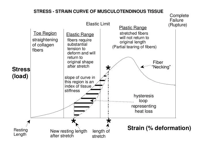
Microtrauma. Microtrauma ( “overuse syndromes” “cumulative cell-matrix adaptive response” ) Repetitive maximal or submaximal stress (movement in the “elastic region”) causes: pathological microscopic tears or lesions r inflammation

E N D
Microtrauma • Microtrauma ( “overuse syndromes” “cumulative cell-matrix adaptive response” ) • Repetitive maximal or submaximal stress (movement in the “elastic region”) causes: • pathological microscopic tears or lesions r inflammation • overwhelming of cell metabolism r can’t maintain structural integrity by u tissue synthesis • tissues that are formed in response to overload stimulus are of inferior quality (scar tissue) • Responsible for 30% - 50% of sports injuries (Herring & Nilson, 1987) • These types of injuries are very “individualistic” in nature • Structures most often affected in microtraumatic syndromes: • bursa - synovial fluid sacs that d friction between: skin-bone, tendon-bone, muscle-muscle • tendon sheaths (paratenon) - fat & areolar tissue - “extended bursa” surrounding tendon • bones (stress fractures) • muscle (delayed onset muscle soreness) • Symptomology progression of microtraumatic overuse syndromes: Phase 1: pain after activity (immediate - 12 hours) which is palpable at injury site Phase 2: pain during & after activity - no significant impairment - eventually resolves Phase 3: pain during & after activity - significant impairment - eventually resolves Phase 4: pain all the time accompanied by significant impairment - no resolution
Theoretical Mechanisms of Delayed Onset Muscle Soreness • Microscopic tears of muscle & connective tissue • evidence: u CPK and myoglobin in the blood • Excess metabolites r osmotic changes in cell r fluid accumulation r pain • Muscle Spasms • evidence: EMG studies (Dr. Bob Arstrong - TAMU muscle biology laboratory) • Eccentric muscle contractions r sarcomere inhomogeneities & sarcolemma disruption • Intracellular [Ca++] u faster than Calcium extrusion mechanisms can pump it out • Disturbance in Ca++ homeostasis reduces ATP production r d [ATP] • u protease & phospholipase activity begin to degrade the myofibril & associated membrane • phagocytes & macrophages invade cell 2-6 hours after the injury - continues for 2-3 days • prostaglandin & histamine r edema and inflammation r stimulation of free nerve endings • cell adapts r future bouts of the same exercise causes less injury
MRI of Delayed Onset Muscle Soreness • Note u signal intensity in entire bicep compared to ticeps
Tendinitis • Tendinitis - inflammation of tendon • term usually used to refer to inflammation of tendon sheath • the correct terms for inflammation of the sheath are "tenosynovitis" & "paratenonitis" • may be accompanied by tissue degeneration (necrosis) and vascular damage • may lead or contribute to complete or partial tendon rupture • chronic tendinitis r fibrinous adhesions r diminished tissue strength & function • although cause & etiology are debatable, recent opinions purport causes to be related to: • the sliding of tendon over other structure (compressive forces) • subjecting the tendon to loads close to tensile strength & exceeding anabolic capabilities • eccentric contractions • negative direction on force velocity curve r plyometric training?????? • exercise r u glucocorticoids and catecholamines r u collagen turnover but tissue quality is inferior • chronic anabolic steroid use r d tissue quality and tensile strength r u incidence of tendon rupture • malnutrition influence: d vitamins A & C, d copper r d collagen synthesis and crosslinking • menopause r d [E2] r d connective tissue elasticity r u tendinitis & other overuse injuries • Tendinosis - degeneration & deleterious changes in tendon without inflammation
General Steps in the Healing of Inflammatory Connective Tissue (Tendinitis, Tenosynovitis, etc.) Stage Pathology, Healing, & Objectives Treatment Implications Inflammatory inflammation r u GAG & collagen synthesis Rest (activity cessation) (days 0 - 6) prevent injury to developing vascualture & collagen NSAID’s, oral corticosteroids prevent prolonged course of inflammation low level ROM exercises cryogenic therapy? Fibroplastic u rate of collagen synthesis by fibroblasts low level / low duration exercise Proliferation exercise & stretching r align synthesized collagen cryogenic-thermogenic therapy? (days 5 - 21) ultrasonography? Remodeling u rate of crosslinkage formation & fibril size progression of activity Maturation replacement of initial or inferior tissues (u intensity & duration) ( > 20 days) Note:many body tissues follow this paradigm (different timeframe)
Immobilization vs. Mobilization: A Fine Line • Effects of immobilization on injured tendinous tissue • protein degradation exceeds protein synthesis r net d in collagen quantity • reduction in the number of collagen crosslink bonds • atrophy of tissues at myotendinous (muscle - tendon) junction • Benefits of mobilization (movement) on injured tendinous tissue • u in the cross-sectional area of the healed tendon • improvement in collagen fiber “type” and fiber arrangement • u number of crosslink bonds • improvement in the quality of the ground substance in the tendon
Common Therapies for Tendinitis • NSAID drugs • Literature reviews conclude approximately 73% of studies show NSAID’s to be effective • d healing time • d inflammation • Approximately 50% of those using NSAID drugs will have adverse side effects • Corticosteroids • short term use of oral corticosteroids (Medrol Dosepack, prednisone) appear helpful for • neuritis • paratenonitis • bursitis • corticosteroid injections • will reduce pain but usefulness in d inflammation & d healing time has not been proven • may result in collagen disarray, inferior tissue quality, tendon rupture • possible side effects: tissue atrophy, skin depigmentation, transient elevation of serum glucose • used only after a 6-week trial of rest, NSAID’s, then re-conditioning • used only when site of pain is palpable - avoid injection directly into tendon • allow 2 - 6 weeks after injection before re-conditioning • avoid more than 3 injections • avoid injections just prior to competition (d pain r u likelihood of injury exacerbation) • Surgery - excision / debridement of inflamed tissue - release & repair, etc • replace “bad scar” with “good scar” - not always successful • does not remove inflammation stimulus - problems can re-appear
MRI of Achilles Tendinitis • [1] u signal intensity r inflammation • [2] Calcaneous Note inhomogeneity of tendon at inflammation site
Bone Stress Continuum:Stress Reaction Stress Fracture Stress Fractures • Stress Reaction: • accelerated adaptive bone remodeling • most often asymptomatic • occurs most often in tarsals, metatarsals, femur, & tibia • sometimes seen on X-ray as increased callus deposition • Stress Fracture: • complete defect or “crack” in the bone • easily seen on X-ray
Stress Fractures • Occurs only in race horses, greyhounds, & man • intensity and / or volume of activity is not natural • Most stress fractures heal by themselves without clinical manifestations • Causes: • repetitive torque across a bone • weight bearing impact forces • bone does not adapt as well as muscle r u risk of injury becoming chronic • inability of weak musculature to facilitate shock absorption • Stress fractures u risk of complete fracture at the stress fracture site • Early stage stress fractures (stress reactions) don’t show up well on X-rays • better detected by bone scan - shows up as “hot spots” • Symptoms: • tenderness & pain over the fracture site • pain relieved by rest but becomes progressively more frequent & persistent • overlying soft tissue may exhibit swelling • tuning fork struck and placed over the fracture site will elicit pain • muscle atrophy
Stress Fractures • Types of stress fractures: • Oblique / Transverse • angled to or perpendicular to the long axis of the bone • most common • most dangerous due to the likelihood of bone “displacement” • Longitudinal • Compression • Examples of Stress Fractures: • tibial stress fracture in runners • weight bearing impact related • humerus stress fracture in javelin thrower • related to muscle trying to accelerate a “resisting” bone (torque)
Stress Fracture of Left Tibia Bone Scan Stress Fracture Site "Hot Spot" Corresponding X-ray
Bilateral Stress Fractures of the Ulna in a Weight Lifter "Hot Spots"
Stress Fractures Stress Fracture of 3rd Metatarsal Stress Reaction of Calcaneous in Runner
Healing of Microtraumatic Injuries