Download
1 / 48
500 likes | 954 Views
Crystal-induced arthritis & Osteoarthritis. Ira Targoff, MD Division of Rheumatology. “The Gout” James Gillary, 1799. Gout- an evil demon attack the toe. Hyperuricemia. Hyperuricemia is defined as a serum uric acid >7 mg/dl in men and >6 mg/dl in women

E N D
Crystal-induced arthritis &Osteoarthritis Ira Targoff, MD Division of Rheumatology
“The Gout”James Gillary, 1799 Gout- an evil demon attack the toe
Hyperuricemia Hyperuricemia is defined as a serum uric acid >7 mg/dl in men and >6 mg/dl in women The majority of people with hyperuricemia never develop any clinical problems from it ("asymptomatic hyperuricemia").
Formation of Uric Acid Hypoxanthine Xanthine Uric acid • Catalyzed by XanthineOxidase (XO)
Causes of Hyperuricemia Underexcretion of uric acid (90%) Renal insufficiency Medications (diuretics, cyclosporine, salicylates, ethanbutol, pyrazinamide) Ethanol, organic acids (lactic acidosis, ketoacidosis) Lead nephropathy Overproduction (10%) High cell turnover (myeloproliferation, psoriasis) High purine diet, ethanol Genetic causes (X-linked) 1) Hypoxanthine-guanine phosphorybosyl trnaferase (HGPRT) deficiency 2) 5-phosphryboyl-1-pyrophosphate synthetase (PRPP synthetase) over activity
How to distinguish between overproducers and underexecreters? • 24-hr urine collection for uric acid Uric acid >800 mg/24hrs Overproducer Uric acid <800 mg/24hrs Underexecreter • Spot urine uric acid
The famous Podagra -Sudden onset of severe pain and swelling at night -Usually single joint (85%) or fewer than 4 joints -Polyarticular gout is a later feature. -Fever and leukocystosis -Spontaneous resolution over 1-2 weeks -The red apple is replaced by purple plum with exfoliating skin.
Gout-epidemiology • The prevalence of gout in the adults is 1.4%, and in men over 75 is 7% (UK General Practice Data). • Gout is rare in males under 30 and in women before menopause • The peak age of onset in men is 40-50 and in women is after 60 • There is an association between hyperuricemia and metabolic syndrome (insulin resistance, hypertension, obesity, dyslipidemia)
Mono-sodium urate crystals A. Perpendicular to Polarizer Axis B. Extinction C. Parallel to Polarizer Axis Polarizing Axis
Axis Uric acid crystals parallel to the polarizing axis are yellow, those perpendicular are blue
Mono-sodium urate: Intracellular Intracellular crystals indicate acute flare
Crystal Analysis by Polarized Light Microscopy Presence of Crystals Shape of crystals Needle vs Rhomboid vs Other Birefringence Sign of birefringence Negative birefringence (yellow): MSU Positive birefringence (blue): CPPD Apatite not visible by this method
Gout: X-ray Tophus Rat bite erosions with overhanging edges
Treatment of gout Acute attack Colchicine NSAIDs Steroids (injection or systemic) Uric acid lowering agents Allopurinol: Xanthine oxidase inhibitor Probenecid: Uricosuric; inhibits urate transporter-1 (URAT-1) Febuxostat: Non-purine xanthine oxidase inhibitor Uricase
Indications for uric acid lowering agents • >2 attacks within 1-2 yrs • Renal stones (urate or calcium) • Tophaceous gout • Chronic gouty arthritis with erosive disease • Asymptomatic hyperuricemia >12 mg/dl or 24 hrs urinary excretion >1100 mg (to decrease risk for uratenephrolithiasis)
Calcium Pyrophosphate Dihydrate (CPPD) Deposition Disease “Pseudogout” : acute attacks may be provoked by pamidronate, G-CSF, intra-articularhyaluronic acid, joint lavage or surgery) Pyrophosphate arthropathy “pseudo-osteoarthritis” OA involving the knees, wrists, metacarpophalangeal joints, (particularly the second and third), shoulders and hips. “Pseudo-rheumatoid” polyarthritis (MCPs and wrists) Asymptomatic (most common)
Calcium Pyrophosphate Dihydrate (CPPD) Deposition Disease Associated predisposing conditions include: • Hemochromatosis • Hyperparathyroidism • Hypomagnesemia • Hypophosphatasia • Increasing age
Calcium Pyrophosphate Dihydrate Deposition Wrap around patella “Fuzzy” calcium in the joint space
CPPD: MCPS Squaring off and large hook-like osteophytes on 2nd and 3rd MCPS
CPPD, wrist Joint destruction Calcium Cystic changes
CPPD Shoulder • High riding • Sclerosis of glenohumeral space
Calcium Pyrophosphate Dihydrate Deposition Rhomboid crystals are faintly blue in parallel Axis
Other Synovial Fluid Crystals • Basic Calcium Phosphate; hydroxyapetite • acute calcificperiarthritis • acute arthritis • destructive arthropathy (Milwaukee Shoulder) • Calcium Oxalate • acute arthritis • Lipid • Cholesterol
A heterogeneous group of conditions that lead to joint symptoms and signs which are associated with defective integrity of articular cartilage, in addition to related changes in the underlying bone at the joint margins. American College of Rheumatology Definition of Osteoarthritis
Osteoarthritis (OA) • Fastest growing disease in the US • Most common arthritis • 16 million patients in US alone • Mainly affects 60 years old • 80% of people 75 have OA symptoms
OA - Clinical Features • Asymmetrical arthritis • Primarily affects weight bearing joints (knees, hips) • Most common joints - knees, hips, DIP’s, 1st MCP, 1st CMC • Little morning stiffness (<30min) • Increased Pain with activity
OA - Diagnosis Clinical and radiographic No elevation of sedimentation rate Normal hematocrit and leukocyte count Non-inflammatory joint tap ( usually <2,000) No crystals in synovial fluid
OA of hand Bone on bone loss of joint space PIPs and DIPs 1st Carpal metacarpal at base of thumb Spares MCPs CMC
Histology of OA Loss of articular cartilage and osteophyte formation
Carpometacarpal Joint OA • Loss of joint space • Sclerosis • Subchondral cysts • Subluxation
Osteoarthritis of Left Hip Loss of joint space Sclerosis Osteophytes Normal
OA of knee Osteoarthritis
Pathology of Knee OA Loss of articular cartilage on medial condyle, intercondylar surface and patella
OA Risk factors • Increased age • Positive family history • Obesity
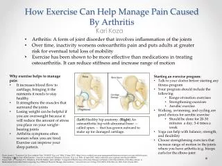
OA - Therapy Patient education Joint safe exercise program Nutritional counseling for obesity Rehabilitation, including joint sparing methods of ADLs, isometric exercises, etc. Nutritional supplements: glucosamine? Pharmacological therapy Joint replacement surgery
OA - Drug Therapy • Simple analgesia • Round the clock acetaminophen • 4000 mg per day (every day, not prn) • Two extra-strength four times a day • Not effective in patients with a “gelling” component (will need NSAID) • Not effective in patients with signifcant morning stiffness • Poor compliance due to large number of pills • NSAIDs: scheduled, once dialy or twice daily have better compliance • Hyaluronic acid injection, steroid injections • Capsacian cream