Download
1 / 19
260 likes | 1.36k Views
Shoulder. Glenohumeral Joint. AP shoulder girdle. Three projections with different positions of the arm will demonstrate the humeral head & neck in different views. AP with arm in external rotation – True AP AP with arm in neutral position

E N D
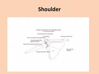
Shoulder Glenohumeral Joint
AP shoulder girdle Three projections with different positions of the arm will demonstrate the humeral head & neck in different views. • AP with arm in external rotation – True AP • AP with arm in neutral position • AP with arm in internal rotation –humerus in lateral
External rotation Greater tubercle (arrow) Lesser tubercle (arrowhead) Neutral rotation Internal rotation
AP with arm in external rotation – True AP Patient & part position • Supine or erect • Rotate patient slightly to place the spine of the scapula approximately parallel with the plane of the cassette • Abduct the arm slightly andthe palm forward to bring the coronal plane of the epicondyles parallel to the cassette
AP with arm in neutral position Patient & part position • Supine or erect • Rotate patient slightly to place the spine of the scapula approximately parallel with the plane of the cassette • Rest the palm of the hand against the thigh to bring the humerus in neutral position • Direct Central ray perpendicular to the cassette over coracoid process.
AP with arm in internal rotation –humerus in lateral Patient & part position • Supine or erect • Rotate patient slightly to place the spine of the scapula approximately parallel with the plane of the cassette • Flex the elbow somewhat and rotate the arm internally and rest the back of the hand on hips to bring the humerus in lateral position • Direct Central ray perpendicular to the cassette over coracoid process.
AP oblique for glenohumeral joint Patient & part position • Supine or erect • Rotate patient about 350 to place the body of the scapula parallel with the plane of the cassette • Abduct the arm slightly in internal rotation • Direct Central ray perpendicular to a point 5 cm medial and 5 cm below superolateral border of the shoulder (over coracoid process).
Shoulder Axial • Supero-inferior • Infero-superior
Shoulder AxialSuperoinferior • Direct the central ray through the shoulder joint with the tube angled 5 -10 degrees towards the elbow
Shoulder AxialSuperoinferior • Patient seated on a chair close to the edge of the table • Raise the arm as close as possible right angles to the body • Lean the patient laterally to bring the axilla over the cassette while elbow rests on the table • Elbow flexed at 900 and hand pronated • Turn the head towards unaffected side
PA oblique (scapula Y) Useful in the evaluation of suspected shoulder dislocations
Supraspinatus “Outlet” • To demonstrate tangentially the coracoacromial arch or outlet to diagnose shoulder impingement • The tangential image is obtained by projecting the x-ray beam under the acromion and AC joint, which defines the superior border of the coracoacromial outlet.
Outlet view – for shoulder impingement RAO/LAO(Modified scapula Y projection) • Patient upright and lateral with affected shoulder to center of the bucky • Rotate patient forward to make body of scapula perpendicular to cassette • Elbow flexed and forearm across the anterior (or posterior for body of scapula) chest • Direct central ray angled 100 down from horizontal through head of humerus
AP axial (Stryker ‘notch’ view) • To demonstrate ‘Hill-Sachs defect’ • Anterior dislocations of the shoulder frequently result in posterior defects involving the posterolateral head of the humerus, called Hill-Sachs defects.
Transthoracic lateral • To demonstrate proximal humerus in a 90 degree projection from the AP projection when trauma exists and the arm cannot be rotated or abducted because of an injury