Download
1 / 22
220 likes | 428 Views
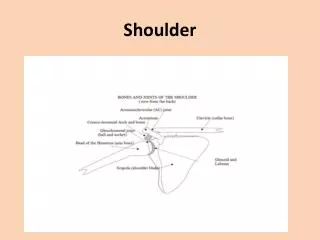
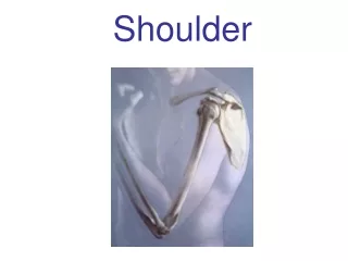
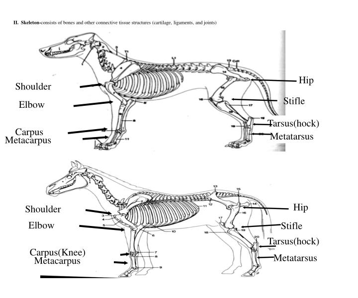
Shoulder. Elbow. Carpus. Metacarpus. II. Skeleton- consists of bones and other connective tissue structures (cartilage, ligaments, and joints). Hip. Stifle. Tarsus(hock). Metatarsus. Hip. Shoulder. Elbow. Stifle. Tarsus(hock). Carpus(Knee). Metatarsus. Metacarpus.

E N D
Shoulder Elbow Carpus Metacarpus II. Skeleton-consists of bones and other connective tissue structures (cartilage, ligaments, and joints) Hip Stifle Tarsus(hock) Metatarsus Hip Shoulder Elbow Stifle Tarsus(hock) Carpus(Knee) Metatarsus Metacarpus
BONES OF THE PELVIC LIMB • The pelvic girdle, or pelvis, of the dog consists: • Two hip bones (Os Coxae): • Each hip boneis formed by the fusion three primary bones and the addition of a fourth in early life • Ilium, which articulates with the sacrum. • Ischium is the most caudal • Pubis is located ventromedial to the Ilium and cranial to the large Obturator foramen. - The small acetabular bone, which helps form the acetabulum, is incorporated with the Ilium, Ischium, and pubis when they fuse (about the third month).
Os Coxae 1- The Ilium It can be divided into: wing body The tuber Coxae The tuber Sacrale, The external or gluteal surface The internal or sacropelvic surface
2- Ischium • tuberosity • body • table • ramus. • 3- The pubis • body • two rami.
The acetabulum • a cavity that receives the head of the femur. • Its articular surface is semilunar and is composed of parts of the Ilium, Ischium, and pubis and the acetabular bone in young animals. • The circumference of the articular surface is broken at the caudomedial part by the acetabular notch.
The pelvic canal • short ventrally but long dorsally Its lateral wall is composed of the Ilium, Ischium, and pubis. • The pelvic inlet is limited laterally and ventrally by the Arcuate line. • Its dorsal boundary is the promontory ofthe sacrum. • The pelvic outlet is bounded ventrally by the Ischiatic arch • Mid-dorsally by the first caudal vertebra, and laterally by the superficial gluteal muscle and the sacrotuberous ligament.
The femur: • is a typical long bone with a cylindrical body and two expanded extremities. • Tibia • Fibula
Tarsal Bones • The tarsus between the metatarsals and the leg, is composed of seven tarsal bones the hock • The bones are arranged in three irregular rows. • The proximal row is composed of a long, laterally located calcaneus and a shorter, medially located talus. • Distal Raw: 1st , 2nd , 3rd and 4th tarsal • Central tarsal bone
Metatarsal Bones • The metatarsal bones resemble the metacarpal bones except for the first, which may be divided, rudimentary, or absent.
Phalanges • Those of the hind paw, or pes, are similar to those of the forepaw, or manus. • The first digit, or hallux, is frequently absent. • When present, it is called a dew claw and may vary from • *- a fully developed digit articulating with a normal first metatarsal bone • *- to a vestigial structure composed only of a terminal phalanx.