Download
1 / 21
210 likes | 494 Views
HS130: Anatomy and Physiology II. Unit 7 Seminar: The Urinary System Dr. Daudi K. Langat Adjunct Professor, Kaplan University. Welcome to today’s Seminar! We will begin at the top of the hour. Housekeeping issues (Unit 9 Writing Assignment).

E N D
HS130: Anatomy and Physiology II Unit 7 Seminar: The Urinary System Dr. Daudi K. Langat Adjunct Professor, Kaplan University Welcome to today’s Seminar! We will begin at the top of the hour.
Housekeeping issues (Unit 9 Writing Assignment) • A reminder that the Unit 9 Assignment due date is coming up soon (it is worth 100 points!) • You will continue with your voyage. Your “Fantastic Voyage!” is continuing inside a 55 year old man eating a hamburger, french fries and a root beer. You go through the following systems: • Digestive tract to review the digestion to completion • Absorption at the distal ilieum to the superior mesenteric Vein. • Urinary system, to the left kidney (trace your path from the superior mesenteric vein to the left renal artery, via the hepatic portal vein, heart and lungs • Go into the nephron and follow the urinary pathway to the outside • Wrap up your report by explaining the integration of the body systems in maintaining homeostasis. You must include at least the circulatory, digestive and urinary systems in your explanation. • References should be in APA format. Review the other PowerPoint entitled “APA Style Formatting”for more information.
Seminar Topic: This week we will discuss the major components of the urinary system and the formation of urine. Chapter 17The Urinary System
KIDNEYS • Location—under back muscles, behind parietal peritoneum, just above waistline; right kidney usually a little lower than left.
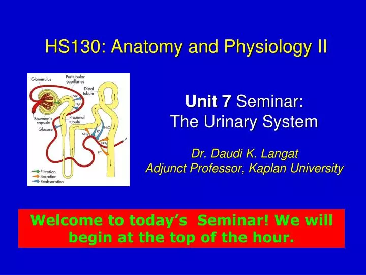
Kidney Internal structure • Cortex —outer layer of kidney substance • Medulla—inner portion of kidney • Pyramids —triangular divisions of medulla • Papilla —narrow, innermost end of pyramid • Pelvis —expansion of upper end of ureter; lies inside kidney • Calyces —divisions of renal pelvis
KIDNEYS • Microscopic structure—nephrons are microscopic units of kidneys; consist of the following (Figure 17-3): • Renal corpuscle • Bowman’s capsule —the cup-shaped top • Glomerulus —network of blood capillaries surrounded by Bowman’s capsule • Renal tubule • Proximal convoluted tubule —first segment • Loop of Henle—extension of proximal tubule; consists of descending limb, loop, and ascending limb • Distal convoluted tubule —extension of ascending limb of loop of Henle • Collecting tubule —straight extension of distal tubule
KIDNEYS • Functions • Excretes toxins and nitrogenous wastes • Regulates levels of many chemicals in blood • Maintains water balance • Helps regulate blood pressure via secretion of renin
FORMATION OF URINE • Occurs by a series of three processes that take place in successive parts of nephron • Filtration —goes on continually in renal corpuscles; glomerular blood pressure causes water and dissolved substances to filter out of glomeruli into Bowman’s capsule; normal glomerular filtration rate 125 mL per minute • Reabsorption —movement of substances out of renal tubules into blood in peritubular capillaries; water, nutrients, and ions are reabsorbed; water is reabsorbed by osmosis from proximal tubules • Secretion —movement of substances into urine in the distal and collecting tubules from blood in peritubular capillaries; hydrogen ions, potassium ions, and certain drugs are secreted by active transport; ammonia is secreted by diffusion • Control of urine volume —mainly by posterior pituitary hormone’s ADH, which decreases it
URETERS • Structure —narrow, long tubes with expanded upper end (renal pelvis) located inside kidney and lined with mucous membrane • Function—drain urine from renal pelvis to urinary bladder
URINARY BLADDER • Structure (Figure 17-7) • Elastic muscular organ, capable of great expansion • Lined with mucous membrane arranged in rugae, as is stomach mucosa • Functions • Storage of urine before voiding • Voiding
URETHRA • Structure • Narrow tube from urinary bladder to exterior • Lined with mucous membrane • Opening of urethra to the exterior called urinary meatus • Functions • Passage of urine from bladder to exterior of the body • Passage of male reproductive fluid (semen) from the body
MICTURITION • Passage of urine from body (also called urination or voiding) • Regulatory sphincters • Internal urethral sphincter (involuntary) • External urethral sphincter (voluntary) • Bladder wall permits storage of urine with little increase in pressure • Emptying reflex • Initiated by stretch reflex in bladder wall • Bladder wall contracts • Internal sphincter relaxes • External sphincter relaxes, and urination occurs
PROBLEMS WITH MICTURITION • Urinary retention —urine produced but not voided • Urinary suppression —no urine produced but bladder is normal • Incontinence —urine is voided involuntarily • May be caused by spinal injury or stroke • Retention of urine may cause cystitis • Cystitis —bladder infection • Overactive bladder —need for frequent urination • Called interstitial cystitis • Amounts voided are small • Extreme urgency and pain are common
Major Disorders of the Urinary System • Hydronephrosis • Calculi (stones) • Neurogenic bladder • Tumors • Urethritis • Infection • Cystitis • Pyelonephritis • Renal Failure
Course Questions, Problems & Issues: Please post in “Instructor Questions” in the class GOODNIGHT EVERYBODY!!