Download
1 / 21
210 likes | 307 Views
Explore microscopic eukaryotes like Paramecium and Amoeba, along with bacterial slides of Micrococcus and Bacillus species. Practice staining techniques and bacterial growth analysis in this informative microbiology lab session.

E N D
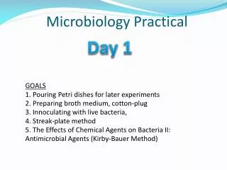
Microbiology Practical I The following slides were contributed by your fellow BIO 251 students… Use side “” key to view slides…
Lab 1: Eukaryotes and Microscopy Know the parts of the microscope…
Lab 1: Eukaryotes and Microscopy Paramecium caudatum
Lab 1: Eukaryotes and Microscopy Euglena gracilis
Lab 1: Eukaryotes and Microscopy Amoeba proteus
Lab 1: Eukaryotes and Microscopy Fungus – Saccharomyces cerevisiae
Lab 1: Eukaryotes and Microscopy Cyanobacteria ~ Nostoc
~Bacteria slides~ Micrococcus luteus
~Bacteria Slides~ Bacillus subtilis
~Bacteria Slides~ Bacillus anthracis
~Bacteria Slides~ Rhodospirillum rubrum Sorry not in focus…
~Bacteria Slides~ See other file for Gram Stain, Endospore, Acidfast, and Capsule Staining… [ Adobe Acrobat of Gram and Endospore ] Some Endospore Staining ~
Streak Plate: Know colony characteristics (color, elevation, margin, etc.)
Spread Plate: Mixed – Bacillus subtilis & Serratia marcescens
Practical I Unknown Tests: ~ Simple Stain ~ Gram Stain ~ Endospore Stain ~ Acid-Fast Stain (Demo) ~ Capsule Stain (Demo) ~ Motility (Hanging Drop, Wet Mount, Soft Agar Deep) ~ Obligate Aerobe, Facultative Anaerobe, Aerotolerant Anaerobes, Microaerophiles (Gas Pak, Eugon Agar, Thioglycollate Broth)
Bacterial Growth Curve: Make sure you know how to: Use a spectrophotometer to measure absorbance. Produce a log graph of the calculations. Calculate generation time from a growth curve.