Download
1 / 61
620 likes | 740 Views
Learn about the types, causes, and treatment of scoliosis in children, including infantile, juvenile, and adolescent idiopathic scoliosis. Discover the different types of scoliotic curves and how to recognize and manage this condition effectively.

E N D
The Scoliosis Dr.Mouayad Kazem Damascus Hospital 27/4/2006
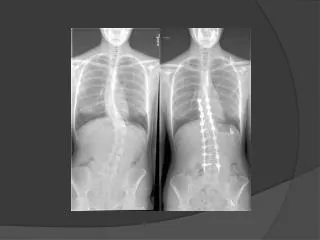
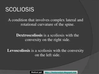
What is the Scoliosis ? • Lateral deviation of the vertebral line of the spine (measured > 10°) • Abnormal movement in 3 planes : • Intervertebral extension • Lateral intervertebral tilting • Rotary component
Classification • Idiopathic : • 80% • Any age – peaks in (1year _ 5-6 years _ 11 year) • Secondary : • Congenital • Neuromuscular • Hysterical
Idiopathic Scoliosis • Infantile Idiopathic Scoliosis • Birth – 3 years old • Juvenile Idiopathic Scoliosis • 4 – 10 years old • Adolescent Idiopathic Scoliosis • 11 year – skeletal maturity
Terms describing different types of Scoliotic curves • Structural curve • Nonstructural curve • Primary curve • Compensatory curve • lordoscoliosis • Kyphoscoliosis
Terms describing different types of Scoliotic curves • Cervicothoracic curve • Apex at C7 – T1 • Thoracic curve • Apex at T2 – T11 • Thoracolumbar curve • Apex at T12 – L1 or T12-L1 interspace • Lumbar curve • Apex at L1 – L4 • Lumbosacral curve • Apex at L5 or below
Terms describing different types of Scoliotic curves • Double curves • Double major curves • Double thoracic curves
Infantile Idiopathic Scoliosis • Birth – 3 years (most in first 6 months) • Left curve 90% • ♂>♀ ( 3 : 2 ) • 90% = self limited • Double major curves → severe deformity • Right thoracic curves + ♀→ bad prognosis.
Infantile Idiopathic Scoliosis • Clinical examination : • The spine , neurologic , associated deformities • Xray evaluation : • Cobb angle • RVAD • Phase of the rib head
Infantile Idiopathic Scoliosis • Rib Vertebral Angle Differencs ( RVAD) RVAD = RVA Concave – RVA Convex RVAD > 20° = progressive RVAD < 20° = self limited
Infantile Idiopathic Scoliosis • Phase of the rib head
Juvenile Idiopathic scoliosis • 12-21% • ♀< ♂(2:1→4:1) • Average age of Diagnosis : • ♀= 7 years • ♂ = 5 years • 70% progress → require treatment • Curve type resemble AIS : Right thoracic – DM
Juvenile Idiopathic scoliosis • Mehta classification : • Late-resolving idiopathic scoliosis • Benign progressive infantile idiopathic scoliosis • syndromic scoliosis • Syringomelic scoliosis • Early detected AIS
Juvenile Idiopathic scoliosis • Clinical examination : • Spinal deformities • Midline dimples • Hairy patches • Neurologic signs • Loss of abdominal reflexes • Absent gag reflex
Juvenile Idiopathic scoliosis • Investigations : • MRI : all of the patients <normal neurologics> Or : in patients <10 year + curve > 20° • Xray : • Cobb angle • RVAD : no benefit in prognosis But : used in predicting response to brace treatment
♂ + Left curve + cobb> 45° + kyphosis < 20° ↓ Progressive Juvenile Idiopathic scoliosis
Adolescent Idiopathic Scoliosis - AIS • ♀>♂(curve>45°= 9:1) • Discovered in school screening tests • No complaints ( cases of pain ) • AIS is a diagnosis of clinical and radiographic exclusion . • Left thoracic curves → syringomyelia
AIS – Before Skeletal Maturity • Factors predict progression : • Sex : Females • Remaining growth Assessment by : • Menarchal status ♀ • Risser sign • Tanner index • Peak Height Velocity PHV
AIS – Before Skeletal Maturity • Risser sign
AIS – Before Skeletal Maturity Tanner stages - Girls
AIS – Before Skeletal Maturity Tanner stages - Boys
AIS – Before Skeletal Maturity • Factors predict progression (cont.): • Curve size : • Risser 0 + premenarch + curve > 20° → high risk of progression • Curve pattern : • DM – Thoracic curves = high risk • Thoracolumbar • Lumbar = least risk
AIS – After Skeletal Maturity • Curves < 30° = unlikely to progress • Curves > 50° = worsen Thoracic curves = 1° / year • Mortality rate is the same • Infantile-Juvenile idiopathic scoliosis = ↑mortality Respiratory failure – cardiovascular disease • Chronic back pain • Lumbar osteoarthritis
Scoliosis screening • signs • Shoulder asymmetry • Unequal scapular prominence • Elevated or prominent hip • Greater space arm-body on one side • Head not centered over the pelvis • Adam forward bending test + • Scoliometer Recommendation for orthopedic referral = 5° trunk rotation
AIS - Etiology • Neurologic dysfunction • Vestibular-eye-proprioceptive systems • ↓ response to vibratory stimuli • Melatonin deficiency • Connective tissue abnormalities: • Difference in collagen • Ligamentum flavum • Paravertebral musculature • ↑ platelet calmodulin level • Genetic factors
AIS - Pathophysiology • Changes are greatest at apex and diminish toward ends • In structural scoliosis : rotation of the vertebral body is to the convexity and the spinous process is to the concavity . • The scoliotic portion of the spine is lordotic in the sagittal plane
AIS - Patient evaluation • Complaint : • Body deformity • Pain : age>15 - risser≥ 2 – postmenarch – history of injury • Most common causes are : • Spondylolysis • Spondylolisthesis • Scheuermann’s kyphosis • Less common causes are: • Spinal cord syrinx • Disk herniation • Tethered spinal cord • Tumor • Respiratory symptoms • Neurologic deficit
AIS - Physical examination • Inspection : • Skin : midline hemangiomas – hair tufts – lumbar dimpling • Body asymmetry • Palpation : • Absence of a spinous process (spina bifida occulta) • Adam’s forward-bending test • Spinal balance
AIS - Physical examination Trunk balance Plumb line
AIS - Physical examination • Neurologic examination : • Patients reflexes • Superficial abdominal reflexes • Muscle test : • Range of motion • Hands and feet • Callus – nail bed irregularities.
AIS - Image study • Plain Xray : • Posteroanterior view (less radiation exposure) • Curve type and size • Trunk and vertebral balance • Skeletal maturity • Pelvic tilt • Appropriate fusion levels • Lateral view : • Lordoscoliosis • Hyper – hypo kyphosis • Spondylolisis - Spondylolisthesis
AIS - Image study • Plain Xray cont. • Bending view : • Curve flexibility • Surgical-nonsurgical indication • Stagnara view Stagnara view
AIS - Image study Curve size: Cobb angle
AIS - Image study Vertebral rotation
Sagittal balance on plain radiograph AIS - Image study
AIS - Image study • Dorsal kyphosis : • Apex at T4-T7 • Size 20-45° • Lumbar lordosis: • Apex at L3-L4 • Size 50-65° • 20% at L4-L5 • 40% at L5-S1 • Lumbar disks = 47° • vertebral bodies = 12° Balanced SVA = lumbar lordosis > thoracic kyphosis 20-30°
AIS - Curve types Ponseti – Friedman classification • Single major lumbar curve • Single major thoracolumbar curve • Combined thoracic and lumbar curves • Single major thoracic curve • Single major high thoracic curve • Double major thoracic curve
AIS – Lenk classification It’s a trial system combining curve type , the lumbar spine modifier and thoracic sagittal modifier eg. 1AN There are 42 different classifications
Scoliosis - Treatment Infantile idiopathic scoliosis: • Cobb <25° + RVAD <20 °→ observation • 5-10° increment of cobb/RVAD → treatment • Methods: • Serial casting • Milwaukee brace
Scoliosis - Treatment Juvenile idiopathic scoliosis • Curve <20° → observation • Curve >25° + flexible → Milwaukee brace • curve <35° + RVAD <20° = excellent prognosis • Curve >45° + RVAD >20° = poor prognosis • Curve >40-50° → Surgical treatment • Depends on : • Age • Spinal growth remaining < essential > • Expected loss of spinal growth • The aim is to halt progression of scoliotic curve. • Crankshaft phenomenon
Treatment – Juvenaile IS Surgical methods : • Instrumentation without fusion or with limited fusion • Hooks at the end vertebrae with rod • The rod is lengthened every 6-12 months • The same with localised fusion at the apex • Lessens curve progression • Luque rod and wires without posterior fusion • No need to lengthen the instrument • No need for external fixation • The same with anterior fusion at the apex