Download
1 / 31
310 likes | 647 Views
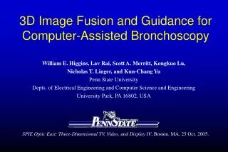
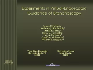
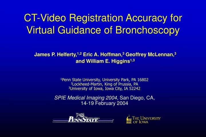
CT-Video Registration Accuracy for Virtual Guidance of Bronchoscopy. James P. Helferty, 1,2 Eric A. Hoffman, 3 Geoffrey McLennan, 3 and William E. Higgins 1,3. 1 Penn State University, University Park, PA 16802 2 Lockheed-Martin, King of Prussia, PA 3 University of Iowa, Iowa City, IA 52242

E N D
CT-Video Registration Accuracy for Virtual Guidance of Bronchoscopy James P. Helferty,1,2 Eric A. Hoffman,3Geoffrey McLennan,3andWilliam E. Higgins1,3 1Penn State University, University Park, PA 16802 2Lockheed-Martin, King of Prussia, PA 3University of Iowa, Iowa City, IA 52242 SPIE Medical Imaging 2004, San Diego, CA, 14-19 February 2004
CT-Guided Bronchoscopy for Lung Cancer Staging ROI seen in CT but not in video CT Scan of Chest • Bronchoscopic biopsy critical for staging. • Physicians make errors when maneuvering bronchoscope to a biopsy site. • Lymph nodes are hidden from endoscopic video, but visible in 3D CT analysis exploit CT using image guidance • CT-guidance of bronchoscopy reduce errors, improve biopsy success rate Videoendoscopy Inside airways Matching Video and CT Rendering
No device needed Image-Guided Bronchoscopy Systems Show potential, but recently proposed systems have limitations: • McAdams et al.(AJR 1998) and Hopper et al.(Radiology 2001) • Virtual bronchoscopy for lymph-node biopsy, but no live guidance. • Solomon et al. (Chest 2000) – E/M sensor attached to scope • limited planning, many potential errors, limited guidance • Bricault et al. (IEEE-TMI 1998) –no device needed • Registered videobronchoscopy to CT, but no live guidance. • Mori et al. (SPIE Med. Imaging 2001, 2002) – no device needed • Registered videobronchoscopy to CT and tracked video. • Efforts not interactive: >20 sec to process each video frame.
PC Enclosure AVI File RGB,Sync,Video Video Stream Matrox Cable Video Capture Main Thread Video Tracking OpenGL Rendering Worker Thread Mutual Information Dual CPU System Scope Monitor Scope Processor Rendered Image Light Source Endoscope Polygons, Viewpoint Image Matrox PCI card Video AGP card Our Group’sImage-Guided Bronchoscopy System Software written in Visual C++.
System Processing Flow Data Sources 3D CT Scan Bronchoscope Stage 1: 3D CT Assessment and Planning • Segment 3D Airway Tree • Calculate Centerline Paths • Define Target ROI biopsy sites Stage 2: Live Bronchoscopy • Capture Endoscopic Video • Correct Video’s Barrel Distortion • Track/Register Video and Virtual CT • Map Target ROIs on Video Image Processing HTML Multimedia Case Study Site List Segmented Airway Tree Centerline Paths Screen Snapshots Recorded Movies Physician Notes See: Helferty et al., SPIE Med. Imaging 2001; Swift et al., Comp. Med. Imag. Graph. 2002.
Display during Stage-2 Bronchoscopy Case h005_512_85. Root site = (253,217,0), seger = (RegGrow, no filter), ROI #2 considered (Blue)
Key Step: CT-Video Registration Stage 2: CT-Guided Bronchoscopy Protocol • Provide Virtual-World CT rendering ICT • Move bronchoscope “close” to ICT target view IV • Register Virtual World to target view IV • Go to Step 1 unless biopsy site reached Endoluminal 3D CT rendering Live video from bronchoscope
Optimal CT rendering IV = Target video co ICT ct IV CT-Video Registration Problem:Viewpoints 6-parameter viewpoint 3D position 3-angle direction Standard camera direction matrix
CT-Video Registration Problem:Optimization Problem h(V), h(CT) – entropies based on image histograms (PDFs) c ICT ci – starting point for Normalized Mutual Information (NMI): NMI Optimization: Ref: Studolme et al., Pattern Recognition, 1/99.
CT-Video Registration Problem:Optimization Algorithms Tested • Steepest Ascent • Nelder-Meade Simplex • Simulated Annealing
co ICT needle position for bronchoscope ( ) “needle” position for optimal CT view ( ) IV CT-Video Registration Problem: Error Measures for Tests Position error Angle error Needle error where: po no
co ICT errors for acceptable registrations Registration Protocol for Tests IV • Target video frame: View to optimize: 2. Registration process: • Fix 5 parameters of ICT’s viewpoint to IV’s true viewpoint: -10 mm < DX, DY, DZ < 10 mm -20o < Da , Db , Dg < 20o • Initialize ICT’s remaining parameter away from true value • Run NMI optimization until convergence • Measure errors
IV • Target video frame: -- known fixed virtual CT view • View to optimize: -- based on SAME 3D CT image as 3. Test each optimization algorithms: stepwise, simplex, annealing IV co ICT Test #1: Performance of Optimization Algorithms(a) Eliminate video and CT source differences (b) Measure registration error precisely
co co co co ICT ICT ICT ICT Test #1 -- Performance of Optimization Algorithms Example Registrations • Test “video” view IV • “Good” simplex result (DX=8mm) • “Poor” annealing result (DY=10mm) • “Poor” annealing result (D yaw = 20o)
na= 5o Threshold for acceptable angle errorna Test #1 -- Performance of Optimization Algorithms Example Error Plot (na) Initial Db (yaw) varied. Other 5 parameters of ICT‘sviewpointc start at “true” values
ROI 3 Test #2: Impact of Airway Morphology -- 6 Test ROIs (a), (b) proximal and distal trachea (c), (d) proximal and distal right main bronchus (e), (f) proximal and distal left main bronchus Run Simplex Algorithm
Test #2: Impact of Airway Morphology Ranges of Starting Points that result in acceptable registrations
CT V CT IV IV ICT Iv ROI Grp 3 co co ICT ICT Compare final registered result to Test #3: Registering CT to Real Video * 6 Matching Test Pairs Compare final registered result to
Test #3: Registering CT to Real Video * ROI-3 Pair Roll g DZ
Test #3: Registering CT to Real Video * Summary over 6 ROI Pairs Ranges of Starting Points that result in acceptable registrations
co TLC ICT ICT Test #4: Sensitivity to Different Lung Capacities * CT scan – done at full inspiration (TLC) * Bronchoscopy – done with chest nearly deflated (FRC) FRC • Target “video” frame: = -- known fixed CT view (from FRC CT volume) • View to optimize: -- CT view from TLC CT volume • Run Simplex optimization algorithm: Compare final result to previously matched result IV ICT co ICT
FRC IV = ICT ROI Pair 2 TLC ICT Test #4: Sensitivity to Different Lung Capacities * 3 TLC/FRC Matching Pairs (Pig data; volume controlled) TLC FRC TLC FRC TLC FRC
Test #4: Sensitivity to Different Lung Capacities * ROI Pair #2 (Pig data; volume controlled) DZ
Test #4: Sensitivity to Different Lung Capacities * 3 TLC/FRC Matching Pairs (Pig data; volume controlled) Ranges of Starting Points that result in acceptable registrations
Discussion • Method successful and runs in near real-time (5 sec per registration). • Good airway segmentation and video/CT “camera” calibration important. • Registration successful: a. over a wide range of anatomy b. Independent of lung volume c. +/- 8-10 mm position deviations, +/-15-20o direction deviation • Head toward continuous video tracking and CT-video registration. Helferty et al. SPIE Med. Imaging 2003 Acknowledgements This work was partially supported by NIH grants #CA074325 and CA091534, and by the Olympus Corporation.
Steepest Ascent Algorithm Also tested Nelder-Meade Simplex and Simulated Annealing
IV IV co ICT Test #2: Impact of Airway Morphology Consider 6 Varied Airway Locations (ROIs) • Target video frame: -- a known fixed virtual CT view • View to optimize: -- based on SAME 3D CT image as 3. Run Simplex optimization algorithm.
V V ICT ICT co co ICT ICT Test #3: Registering CT to Real Video • Target video frame: -- known fixed video frame; have matching • View to optimize: -- from corresponding CT image 3. Run Simplex optimization algorithm: • Fix 5 parameters of ICT’s viewpoint to IV’s true viewpoint • Run optimization • Compare final registered result to 4. Test on three target “video/CT” matching pairs IV
co TLC ICT ICT Test #4: Sensitivity to Different Lung Capacities * CT scan – done at full inspiration (TLC) * Bronchoscopy – done with chest nearly deflated (FRC) FRC • Target “video” frame: = -- known fixed CT view (from FRC CT volume) • View to optimize: -- CT view from TLC CT volume 3. Run Simplex optimization algorithm: • Fix 5 parameters of ICT’s viewpoint to IV’s true viewpoint • Run optimization • Compare final result to previously matched result 4. Test on three “FRC/TLC” matching pairs IV ICT co ICT