Download
1 / 15
150 likes | 154 Views
Learn about the various volumes and capacities of the lungs, gas exchange in the alveoli, and factors affecting respiratory passageways.

E N D
Total lung capacity at maximum inflation Variation in lung with normal, quiet breathing Minimal lung volume (residual volume) at maximum deflation Volume of lungs at end of normal inspiration (average 2,200 ml) Volume of lungs at end of normal inspiration (average 2,200 ml) Difference between end-expiratory and end-inspiratory volume equals tidal volume (average 500 ml) Fig. 12-14a, p. 378
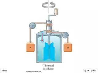
Floating drum Air Recording paper advancing with time Water Expired air Spirogram Inspired air Fig. 12-15, p. 379
TV = Tidal volume (500ml) IRV = Inspiratory reserve volume (3,000 ml) IC = Inspiratory capacity (3,500 ml) ERV = Expiratory reserve volume (1,000 ml) RV = Residual volume (1,200 ml) FRC = Functional residual capacity (2,200 ml) VC = Vital capacity (4,500 ml) TLC = Total lung capacity (5,700 ml) Fig. 12-14b, p. 378
Pulmonary volumes Spirometry: -Volumes: -T.V = 500 ml -IRV= 3000 ml - ERV= 1000 ml - R.V = 1200 ml Capacities : - IC = T.V+IRV 3500 ml - FRC = ERV + RV ml2200 - V.C = IRV + TV + ERV 4500 ml - TLC 5700 ml
Determination of FRC: Helium dilution method : 1- a spirometer is filled with air that is mixed with a known concentration of helium 2- the person expires normally then begins breathing from the spirometer 3- helium becomes diluted by the FRC FRC = (CiHe-1 ) * Vi spir. Cf He RV = FRC - ERV .
Fresh air from inspiration Airway dead-space volume (150 ml) 150 Alveolar air “Old” alveolar air that has exchanged O2 and CO2 with the blood Fresh atmospheric air that has not exchanged O2 and CO2 with the blood After inspiration, before expiration The numbers in the figure represent ml of air. Fig. 12-18a, p. 382
500 ml expired to atmosphere 150 150 ml fresh air from dead space (left from preceding inspiration) 350 ml “old” alveolar air 500 ml “old” alveolar air expired 350 ml expired to atmosphere 150 ml remain in dead space 350 150 “Old” alveolar air that has exchanged O2 and CO2 with the blood Fresh atmospheric air that has not exchanged O2 and CO2 with the blood During expiration The numbers in the figure represent ml of air. Fig. 12-18b, p. 382
150 500 ml fresh air enter from atmosphere 350 ml fresh air reach alveoli 150 ml fresh air remain in dead space 500 ml enter alveoli 350 150 ml “old” air from dead space (left from preceding expiration) 350 ml fresh air from atmosphere 150 “Old” alveolar air that has exchanged O2 and CO2 with the blood Fresh atmospheric air that has not exchanged O2 and CO2 with the blood During inspiration The numbers in the figure represent ml of air. Fig. 12-18c, p. 382
Dead space : not used for gas exchange . • Measurement : 1-deep breath of O2. 2- Expiration into nitrogen meter. 3- first portion recorded by the meter comes from D.S the [N2] increases until a plateau is reached D.S occupies 150 in a normal adult. Physiol. D.S = alv. D.S + anatomical dead space
Minute respiratory volume : Total volume of new air moved into respiratory passages each minute MRV=TV * freq. Normal = 500 x 12 = 6L/min (1.5 L/min fatal). ( high value like 200 L/min is fatal).
Alveolar ventilation : rate at which new air reaches these areas (respir. spaces) . (TV – D.S)* freq. = 4.2L/min
Respiratory passageway: • 1-Main resistance to the airflow present in • Large bronchioles and bronchi • 2-Sympathetic system dilate bronchioles • 3-Parasympatheic system constrict bronchioles • 4-Irritation of membrane passageways cause constriction as(smoking, dust, Infection)
5- Histamine and slow reactive substance of anaphylaxis secrete locally by the lungs • By mast cells during allergic reaction as in • Asthma. These cause bronchiolar constriction • 6-Atropine relax respiratory passageway.