Download
1 / 104
1.07k likes | 1.32k Views
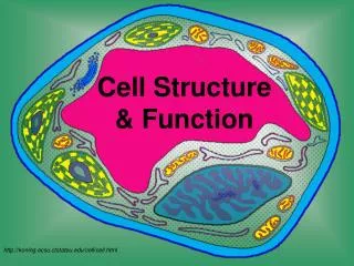
Cells: The Living Units Part A. 3. Cell Theory. The cell is the basic structural and functional unit of life Biochemical activities of cells are dictated by subcellular structure. Structure of a Generalized Cell. Figure 3.2. Plasma Membrane. Important for maintaining homeostasis

E N D
Cells: The Living Units Part A 3
Cell Theory • The cell is the basic structural and functional unit of life • Biochemical activities of cells are dictated by subcellular structure
Structure of a Generalized Cell Figure 3.2
Plasma Membrane • Important for maintaining homeostasis • Separates intracellular fluids from extracellular fluids • Plays a dynamic role in cellular activity • The glycocalyx is a glycoprotein covering on the outside of the cell. It is a marker by which cells recognize one another as well as an ‘adhesive’.
Fluid Mosaic Model • Phospholipid bilayer: a double bilayer of lipids with imbedded, dispersed proteins • Bilayer consists of phospholipids, cholesterol, and glycolipids • Glycolipids are lipids with bound carbohydrate • Phospholipids have hydrophobic and hydrophilic bipoles
Fluid Mosaic Model Figure 3.3
Functions of Membrane Proteins • Transport • Enzymatic activity • Receptors for signal transduction Figure 3.4.1
Functions of Membrane Proteins • Intercellular adhesion • Cell-cell recognition • Attachment to cytoskeleton and extracellular matrix Figure 3.4.2
Plasma Membrane Surfaces • Differ in the kind and amount of lipids they contain • Glycolipids are found only in the outer membrane surface • 20% of all membrane lipid is cholesterol
Membrane Junctions • Tight junction – impermeable junction that encircles the cell • Desmosome – anchoring junction scattered along the sides of cells • Gap junction – a nexus that allows chemical substances to pass between cells
Membrane Junctions: Tight Junction Figure 3.5a
Membrane Junctions: Desmosome Figure 3.5b
Membrane Junctions: Gap Junction Figure 3.5c
Passive Membrane Transport: Diffusion • Simple diffusion – nonpolar and lipid-soluble substances • Diffuse directly through the lipid bilayer • Diffuse through channel proteins
Passive Membrane Transport: Diffusion • Facilitated diffusion • Transport of glucose, amino acids, and ions • Transported substances bind carrier proteins or pass through protein channels
Carriers • Are integral transmembrane proteins • Show specificity for certain polar molecules including sugars and amino acids
Diffusion Through the Plasma Membrane Figure 3.7
Passive Membrane Transport: Osmosis • Occurs when the concentration of a solvent is different on opposite sides of a membrane • Diffusion of water across a semipermeable membrane • Osmolarity – total concentration of solute particles in a solution • Tonicity – how a solution affects cell volume
Effect of Membrane Permeability on Diffusion and Osmosis Figure 3.8a
Effect of Membrane Permeability on Diffusion and Osmosis Figure 3.8b
Passive Membrane Transport: Filtration • The passage of water and solutes through a membrane by hydrostatic pressure • Pressure gradient pushes solute-containing fluid from a higher-pressure area to a lower-pressure area
Effects of Solutions of Varying Tonicity • Isotonic – solutions with the same solute concentration as that of the cytosol • Hypertonic – solutions having greater solute concentration than that of the cytosol • Hypotonic – solutions having lesser solute concentration than that of the cytosol
Active Transport: Sodium-Potassium Pump Extracellular fluid K+ is released and Na+ sites are ready to bind Na+ again; the cycle repeats. 6 Binding of cytoplasmic Na+ to the pump protein stimulates phosphorylation by ATP. 1 Cytoplasm Phosphorylation causes the protein to change its shape. 2 Concentration gradients of K+ and Na+ The shape change expels Na+ to the outside, and extracellular K+ binds. 3 Loss of phosphate restores the original conformation of the pump protein. 5 K+ binding triggers release of the phosphate group. 4 Figure 3.10
Cells: The Living Units Part B 3
Active Transport • Uses ATP to move solutes across a membrane • Requires carrier proteins
Types of Active Transport • Symport system – two substances are moved across a membrane in the same direction • Antiport system – two substances are moved across a membrane in opposite directions
Types of Active Transport • Primary active transport – hydrolysis of ATP phosphorylates the transport protein causing conformational change • Secondary active transport – use of an exchange pump (such as the Na+-K+ pump) indirectly to drive the transport of other solutes
Types of Active Transport Figure 3.11
Vesicular Transport • Transport of large particles and macromolecules across plasma membranes • Exocytosis – moves substance from the cell interior to the extracellular space • Endocytosis – enables large particles and macromolecules to enter the cell
Vesicular Transport • Transcytosis – moving substances into, across, and then out of a cell • Vesicular trafficking – moving substances from one area in the cell to another • Phagocytosis – pseudopods engulf solids and bring them into the cell’s interior
Vesicular Transport • Fluid-phase endocytosis – the plasma membrane infolds, bringing extracellular fluid and solutes into the interior of the cell • Receptor-mediated endocytosis – clathrin-coated pits provide the main route for endocytosis and transcytosis • Non-clathrin-coated vesicles – caveolae that are platforms for a variety of signaling molecules
Exocytosis Figure 3.12a
Clathrin-Mediated Endocytosis Figure 3.13
Membrane Potential • Voltage across a membrane • Resting membrane potential – the point where K+ potential is balanced by the membrane potential • Ranges from –20 to –200 mV • Results from Na+ and K+ concentration gradients across the membrane • Differential permeability of the plasma membrane to Na+ and K+ • Steady state – potential maintained by active transport of ions
Generation and Maintenance of Membrane Potential Figure 3.15
Cytoplasm • Cytoplasm – material between plasma membrane and the nucleus • Cytosol – largely water with dissolved protein, salts, sugars, and other solutes • Cytoplasmic organelles – metabolic machinery of the cell • Inclusions – chemical substances such as glycosomes, glycogen granules, and pigment
Cytoplasmic Organelles • Specialized cellular compartments • Membranous • Mitochondria, peroxisomes, lysosomes, endoplasmic reticulum, and Golgi apparatus • Nonmembranous • Cytoskeleton, centrioles, and ribosomes
Mitochondria • Double membrane structure with shelflike cristae • Provide most of the cell’s ATP via aerobic cellular respiration • Contain their own DNA and RNA
Mitochondria Figure 3.17
Ribosomes • Granules containing protein and rRNA • Site of protein synthesis • Free ribosomes synthesize soluble proteins • Membrane-bound ribosomes synthesize proteins to be incorporated into membranes
Endoplasmic Reticulum (ER) • Interconnected tubes and parallel membranes enclosing cisternae • Continuous with the nuclear membrane • Two varieties – rough ER and smooth ER
Endoplasmic Reticulum (ER) Figure 3.18a and c
Rough (ER) • External surface studded with ribosomes • Manufactures all secreted proteins • Responsible for the synthesis of integral membrane proteins and phospholipids for cell membranes
Smooth ER • Tubules arranged in a looping network • Catalyzes the following reactions in various organs of the body • In the liver – lipid and cholesterol metabolism, breakdown of glycogen and, along with the kidneys, detoxification of drugs • In the testes – synthesis of steroid-based hormones • In the intestinal cells – absorption, synthesis, and transport of fats • In skeletal and cardiac muscle – storage and release of calcium
Cells: The Living Units Part C 3
Golgi Apparatus • Stacked and flattened membranous sacs continuous with ER • Functions in modification, concentration, and packaging of proteins • Transport vessels from the ER fuse with the cis face of the Golgi apparatus • Proteins then pass through the Golgi apparatus to the trans face • Secretory vesicles leave the trans face of the Golgi stack and move to designated parts of the cell
Golgi Apparatus Figure 3.20a
Role of the Golgi Apparatus Figure 3.21