Download
1 / 55
1.12k likes | 3.7k Views
PNEUMONIA. VL – 9 Dec. 24 th 2013 Mohammed El-Khateeb. History of Pneumonia. Described as early as 400 BC by a Greek Physician named Hippocrates. Edwin Klebs was the first to see bacterial infection from a person who died from pneumonia.

E N D
PNEUMONIA VL – 9 Dec. 24th 2013 Mohammed El-Khateeb
History of Pneumonia • Described as early as 400 BC by a Greek Physician named Hippocrates. • Edwin Klebs was the first to see bacterial infection from a person who died from pneumonia. • Described by Sir William Osler over 100 years ago linking the infection to a bacterial cause. • Pneumonia killed a majority of the 50-100 million people that died from the Spanish flu in 1918.
Sir William Osler - - 1892 • "Pneumonia is an infectious disease characterized by inflammation of the lungs and constitutional disturbances of varying intensity. • The fever terminates abruptly by crisis. Secondary infective processes are common. Diplococcus pneumoniae, which is now known as Streptococcus pneumoniae, is invariably found in the diseased lung. • Pneumonia is a self-limited disease and runs its course uninfluenced by medicine."
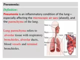
WHAT IS PNEUMONIA Definition: Acute inflammation of lung parenchyma, Inflammatory infiltrate in alveoli (consolidation) It is the infection of one or both of the lungs. Occurs from bacteria, virus, or fungus that is inhaled or gets into the blood stream.
Etiology • Birth – 1 month • Bacterial > Viral • Aspiration of maternal genital organisms • B. Strep • E. coli • 1 to 24 months • Viruses most common • RSV, parainfluenza virus, influenza virus, adeno virus • Apneic episodes • RSV, chlamydia, pertussis
Etiology • 2 to 5 years • Overall rate decreased • Bacterial increase in proportion • S. pneumoniae • Hib sharply decreased after vaccine • Viruses • Influenza A and B, adenovirus • School age and adolescence • M. pneumoniae most common bacterial cause • Peak 10 – 15 years
Viral pneumonia • Gives a pattern of acute injury similar to adult respiratory distress syndrome (ARDS) • Acute inflammatory infiltration less obvious • Viruses recently recognized as important pathogens in CAP due to improved diagnostic tests (PCR) • Cause of 2 - 35% of CAP in adults (more in kids) • Recent emergence of new viral respiratory pathogens
Specific viral pathogens Ruuskanen et al. Viral pneumonia. Lancet. 2011 Apr 9;377(9773):1264-75
The most common causes for viral pneumonia • Influenza • Parainfluenza • Adenovirus • Respiratory syncytial virus (RSV) • appears mostly in children • Cytomegalovirus • In immunocompromised hosts.
Risk factors for viral PNA in adults Marcos MA, Esperatti M, and Torres A. Viral pneumonia. Curr Opin Infect Dis 22:143–147 Elderly: Higher rates of hospitalization and death from viral PNA in persons >60 years of age Chronic Obstructive Pulmonary Disease (COPD) and Asthma: frequently complicated by respiratory viral infections Immunocompromised patients at increased risk Falsey AR, Walsh EE. Viral pneumonia in older adults. Clin Infect Dis. 2006 Feb 15;42(4):518-24
Factors that Contribute to Severe Respiratory Infections Associated with age • Respiratory Factors • Decrease respiratory muscles strength • Decrease protective mucus level • Decrease lung compliance • Decrease level of elastin and collagen in alveolar Ducts • Innate Immunity response • Decreased NK cell cytotoxicity • Decreased NK cell response to IL-2 • Increased level of TNF, IL-1, Il-6 and Il-8 levels
Factors that Contribute to Severe Respiratory Infections Associated with age • Immune Function • Cellular Immunity • Decrease Naïve T cell count • Decrease memory cell count • Decreased T cells proliferation • Imbalance between Th1 and Th2 response • Increased level of inflammatory mediators • Humoral Immunity • Decreased response of B-Cell to new antigens • Increased Autoantibodies
Types of Pneumonia • Bacterial Pneumonia • Viral Pneumonia • Fungal Pneumonia • Parasitic Pneumonia • Atypical Pneumonia • Community-Acquired Pneumonia • Hospital-Acquired Pneumonia • Healthcare-Associated Pneumonia • Ventilator-Associated Pneumonia • Aspiration Pneumonia • Eosinophilic pneumonia • Bronchiolitis obliterans organizing pneumonia
Clinical syndromes • Upper respiratory tract (cold, pharyngitis, bronchitis) • Bronchiolitis: acute inflammatory disorder of small airways • obstruction with air trapping, hyperinflation, wheezing. • Most common < 2 yo • RSV most common, also human metapneumovirus, parainfluenza viruses, influenza A and B viruses, adenoviruses, measles virus, and rhinovirus • Pneumonia • Similar presentation to bacterial PNA Murray and Nadel’s Textbook of Respiratory Medicine 5th Edition
Signs and Symptoms What we will commonly see and hear in the field • Fever • Cough • Cough will bring up Greenish, Yellowish Mucus and possibly hemoptysis • Stabbing Chest pain that worsens with deep respirations • Fatigue • Head Ache • Loss of Appetite • Shortness of Breath • Cyanotic, Sweaty, clammy skin • Rapid Heart Rate • Crackles (Rales)/Wheezing Auscultated • Diminished lung sounds in areas filled with infection
Clinical features • Associated findings • Wheezing, rhinitis, conjunctivitis, pharyngitis • Dehydration • Mental status changes in advanced
Community acquired vs. nosocomial infection • Nosocomial infection: • Often patients in ICU • ↓Local resistance to infection in lungs • Intubation of respiratory tract • Altered normal flora due to antibiotics
Community Acquired Pneumonia • Lower respiratory tract infections are the leading cause of hospitalization for young children worldwide. • Community-acquired pneumonia is a common cause of morbidity and mortality among children in developing countries. • Incidence of CAP in developing countries estimated around150.7million cases/year. • Mortality rate from CAP in developing countries is as high as 2.1 million cases/year. (20% of all mortality cases).
Community Acquired Pneumonia • Viruses have been most commonly associated with CAP in children < 5yr (50-90%). • A limited number of well-defined prospective study of causative agents of CAP, especially in children. • Inconsistent results of the studies.
Viral Pneumonia • Caused by Influenza, parainfluenza, adenovirus, rhinovirus, herpes simplex virus along with several other kinds of viruses. • Antibiotics are not effective in treating viral pneumonia. • It is often treated with antiviral medications along with plenty of fluid and rest • Individuals with suppressed immune systems are most at risk for acquiring this form of pneumonia
Community-Acquired Pneumonia • Community Acquired means that an individual has not been recently hospitalized and has acquired a lung infection • Most commonly caused by streptococcus • Can also be caused by Haemophilus, influenzae, Legionella, mycoplasma, chlamydia, and viruses. • Occurs most commonly in the very young and the very old • Usually starts from an upper respiratory tract infection • S/S usually are that of a flu along with a productive cough with sputum that is rust colored from blood. • Leads to sepsis • Vaccine is available for 23 of the known pneumococcus • Can be treated with antibiotics • Problem with antibiotic resistant strains
Diagnosis in Community Setting From: Pneumonia The Forgotten Killer of Children. Geneva: World Health Organization (WHO)/United Nations Children’s Fund (UNICEF), 2006.
Epidemiology • Incidence of pneumonia decreases as a function of age in children • Seasonal variation • Fall • Parainfluenza virus • Winter • RSV • Bacterial pneumonia (due to indoor crowding) • Spring • Influenza
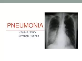
Diagnosis Diagnosed on basis of • Physical examination, • X-Ray findings, and • Laboratory testing
Radiological Examinations • Chest X-ray confirms pneumonia and pleural effusion or empyema • Confluent lobar consolidation is typically in pneumococcal causes • Viral pneumonia- hyperinflation with bilateral interstitial infiltrates • Bronchoscopy, CT scan in malformation or tumors
Pathology • A classical acute inflammatory response • Exudation of fibrin-rich fluid • Neutrophil infiltration • Macrophage infiltration • Resolution • Immune system plays a part antibodies lead to opsonisation, phagocytosis of bacteria • Viral inclusions sometimes seen in epithelial cells
Lab Diagnosis Nasal swab specimens, nasal aspirates, or combined nose and throat swab specimens. Sputum, endotracheal aspirate samples, or BAL Rapid antigen detection, viral culture and PCR methods
Diagnostic Methods • WBC in viral pneumonia are normal or <15,000/ml, with lymphocyte rises; in bacterial WBC>20,000/ml, granulocyte rises • Atypical pneumonia: a higher WBC, ESR and C-reactive protein • PCR test , DNA, RNA, • antibodies tests for the rapid detection of viruses • Serum IgE in recurrent wheezing • Isolation of the bacteria from the blood, pleural fluid or lung • Culture of sputum and susceptibility of the antibiotics • Urinary antigen test positive
Treatment • Treatment depend on the causative agent: • Bacterial Treatment are antibiotics • Viral agents for unusual pneumonia • Varicella pneumonia Acyclovir • RSV Ribavirin, if high risk • HIV Prednisone and zidovudine • CMV Gabcyclovir and Gamma globulin
Complications • Organisation (fibrous scarring) • Abscess • Bronchiectasis • Empyema (pus in the pleural cavity)
Severe Acute Respiratory Syndrome SARS • First identified in Guangdong Province, China • Associated Coronavirus SARS-HCoV • SARS is a form of viral pneumonia where infection encompasses the lower respiratory tract.
HOTEL M IN HONGKONGFebruary 21 Hong Kong 95 Vietnam 37 Ireland 0 Singapore 34 United State 1
Coronavirues • Enveloped • Replicates in cytoplasm of animal cells • Single-strand 30 kb RNA genome • With 5’ cap & poly-A tail • Respiratory, enteric, hepatic, neurological • Cause 30% respiratory infections • Can acquire genes by horizontal transfer and co-infection • Three main classes
Severe Acute Respiratory Syndrome (SARS) • Corona of spikes Made • of S glycoprotein(red) • Cell envelop derived from • Host cell (green) • Core (purplish) M protein • caries the genetic material • (RNA)
The SARS virus has been mutating rapidly in Hong Kong: Mortality rate
SARS CoV - infectivity • Most transmission via close contact with a symptomatic person via large respiratory droplets. Transmission by fomites possible. • Those severely ill more infectious (attack rate of >50% in some hospital staff) • Infectivity increases during second week of illness • Transmission from an asymptomatic person unlikely • May remain infectious up to 10 days once afebrile
Clinical course - triphasic Week 1 • Fever, myalgia, systemic symptoms that improve after a few days Week 2 • Fever returns, oxygen desaturation, CXR worsens Later • 20% get ARDS needing ventilation
Clinical case definition A respiratory illness severe enough for hospitalisation and include a history of: • Fever (> 380C) and • one or more symptoms of respiratory tract illness (cough, difficulty breathing) and • CXR of lung infiltrates consistent with pneumonia or RDS or PM consistent with pneumonia or RDS without an identifiable cause and • No alternative diagnosis to fully explain the illness
SARS - morbidity • Most cases are in healthcare workers caring for SARS patients and close family members of SARS patients • Overall mortality 15% • Mortality increases with age (> 65 years - 50% mortality) • Children seem to develop mild illness
SARS diagnosis • Clinical findings of an atypical pneumonia not attributed to other causes • Exposure to suspect/probable SARS • Or exposure to their respiratory secretions or body
SARS laboratory diagnosis • PCR positive for SARS CoV using validated methods on at least 2 different clinical specimens • Seroconversion(gold standard) (negative antibody test on acute specimen followed by positive test on convalescent sera or > 4 rise in antibody titre between acute and convalescent sera)
SARS - treatment • Supportive, avoid aerosol inducing interventions • Evidence base for anti-viral drugs lacking: (oseltamivir, and intravenous ribavirin). • Antibiotics • Steroids may be helpful • Mechanical ventilation was required in five patients.
Severe Acute Respiratory Syndrome (SARS) Demography Travel 94%
SARS Virus • Survived as long as 24 hours in the environment. • Finding of virus in faeces. • Occasionally linked with pneumonia in humans, specially with immunocompromised. • Can cause severe illness in animals. • Incubation period: 2-7 days
Summary of SARS • Causative agent: Corona virus (Urbani SARS) • Incubation period: 2-7 days (10 days) • Mode of spread: Droplet • Commonest age group: 25-70 years. • Clinical: Fever, respiratory illness, myalgia are common symptoms. Respiratory failure is high. • Adverse outcome: High LDH, high absolute neutrophil count. • Preventive measures are important.
Middle East Respiratory Syndrome Coronavirus (MERS-CoV) • Novel coronavirus that emerged in 2012 • Causes severe acute respiratory illness • First cluster of 2 cases occurred near Amman, Jordan April 2012
MERS-CoV Symptoms • Severe acute respiratory illness: • Fever • Cough • Shortness of breath • Illness onsets were from April 2012 through June 2013 • Some cases have had atypical presentations: • Initially presented with abdominal pain and diarrhea and later developed respiratory complications
MERS-CoV Transmission • Airborne • Incubation period is 10-14 days • The following have been observed: • Transmission between close contacts • Transmission from infected patients to healthcare personnel • Eight clusters of illnesses have been reported by six countries • So far, all cases have a direct or indirect link to one of four countries: Saudi Arabia, Qatar, Jordan, and the United Arab Emirates