Download
1 / 15
150 likes | 219 Views
Explore the intricate details of temporal bone components, facial nerve course, jugular foramen anatomy, and more. Learn about EAC, ossicles, and tympanic membrane structure. Enhance your knowledge of ear anatomy today!

E N D
Sameer Ahmed 9/25/2013 Cummings Ch 127: T-Bone and Ear Anatomy
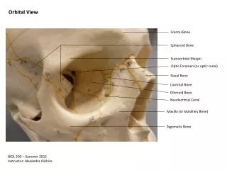
The temporal bone consists of four embryologically distinct components: • Squamous, mastoid, petrous, and tympanic parts • A horizontal ridge known as the temporal line is formed along the most inferior insertion by the temporalis muscle • Aligned with the zygomatic process, • Surface landmark that estimates the location of the middle fossa floor (Tegmen) • “The middle fossa dural plate (MFD) is located on average 5 mm above the LTI (linea tempralis inferior)” • “This study confirms that the mastoid antrum is located 15 mm deep to the lateral surface of the mastoid bone”
Facial Nerve Course • Cisternal segment/intracranial segment • Brainstem to IAC --> 16 -24 mm • Meatal segment → 8mm • Porus acoustics: Medial IAC • Fundus: Lateral IAC • Labyrinthine segment → 5mm • Fundus to geniculate ganglion(1st genu) • Tympanic segment → 8-11mm • Geniculate to 2nd genu • Courses above oval window; dehisc 50% of time • 2nd genu is just anteroinferior to the horizontal SCC • Vertical segment → 10-14mm • 2nd genu to stylomastoid foramen
At the fundus (lateral IAC) • Bill's Bar separates CN 7 anteriorly from SVN • “Jail Bar” • Falciform cr. separates CN 7 from cochlear n.; SVN from IVN • Anatomic relationship changes closer to the brainstem
Jugular foramen: • Pars nervosa: CN X, CN XI, Arnold's nerve, jugular bulb, and posterior meningeal branch of ascending pharyngeal artery • Pars venosa: CN IX, Jacobson nerve, and venous return from inferior petrosal sinus • Inferior limit for a translab approach to IAC • Cochlear aqueduct • The cochlear aqueduct eventually opens into the scala tympani at the cochlear base • Keel: Ridge of bone between Jugular bulb and ICA
EAC: • Lateral cartilaginous: 1/3rd • Lateral canal skin thicker, more sebaceous units • Medial bony: 2/3rd • Think skin; continuous with TM epithelium • Bony-cartilaginous junction in EAC • Site of granulation tissue in malignant OE • Routes of tumor/infxn spread • Foramen of Hushke • Incomplete ossification of bony anterior EAC; medial • Fissures of Santorini • Defects in cartilaginous EAC
1st and 2nd branchial arches → external ear • 6 Hillocks of His • 1st branchial arch: 1-3 hillocks (tragus, superior helix) • 2nd branchial arch: 4-6 hillocks (antihelix, antitragus, lobule, and inferior helix) • External ear blood supply • Posterior auricular & Superficial temporal art.
Tympanic Membrane • Outer layer: epidermal/squamous (ectoderm) • Middle layer: fibrous (mesoderm) • Can be subdivided into radial outer and circular inner • Inner layer: mucosal (endoderm) • Fibrous annulus: thickened pars tensa forming a fibrous outler ring for the attachement to the T-Bone; lies within tympanic sulcus except superiorly where it is deficient at the Notch of Rivinus • Pars flaccida = Shrapnell's Membrane
Eustachain Tube • Medial 1/3rd → Bony • Lateral 2/3rd → Cartilaginous • Collapsed at rest • Tensor veli palatini intermittenly enlarges ET during yawning or swallowing • Bony cartilaginous jnxn of ET is the narrowest portion of the ET • Carotid artery is just medial to ET
Middle Ear • Relative to tympanic annulus • Epitympanum: above annulus • Mesotympanum: confined by the annulus • Hypotympanum: below annulus • Mestoympanum • Anterior limit: ET • Posterior limit: Facial nerve • Medial wall: Cochlear promontory • Postero-superior: Oval window • Postero-inferior: Round window • Sinus tympani: posterior to both windows and medial to vertical division of FN • Pyramidal eminence: anterior to 2nd genu
Hypotympanum • Limited inferiorly by the jugular bulb • Epitymapnum • Superior-medial wall of bony EAC (scutum) forms the lateral wall of the epitympanum • Epitympanum divided into 3 spaces: • Prussak's space, just medial to pars flaccida and lateral to the head and neck of the malleus; • The compartment anterior to the malleus • The posterior compartment, which communicates with the antrum • Attic Cholesteatomas can spread postero-superiorly into the antrum or postero-inferiorly into posterior mesotympanum
Ossicles • Malleus • Manubrium (handle; tip is the umbo) • Lateral/short process • Anterior process • Head • Neck • Tensor tympani attaches to malleus neck and manubrium by a tendon originating from the cochleariform process
Incus • Body • Short process • Long process • Lenticular process • Diarthrodial joints (for malleus-incus and incus-stapes connections)
Stapes • Head (capitulum) • Anterior and posterior crus • Footplate (base) • Stapes footplate attaches to bony margin of oval window via an annular ligament (syndesmosis joint)