Download
1 / 23
240 likes | 685 Views
Corrosive injury. 報告人 : R3 張淳翔. Introduction. Two groups: Pediatric: <5 y/o, accidental ingestion In Taiwan: alkaline oil Adult: Suicidal attempts, intentional More serious corrosive properties In Taiwan: 無煙鹽酸及通樂 (Alkalis) 、魔術靈、漂白水 … Outcome: Caustic properties

E N D
Corrosive injury 報告人: R3 張淳翔
Introduction • Two groups: • Pediatric: • <5 y/o, accidental ingestion • In Taiwan: alkaline oil • Adult: • Suicidal attempts, intentional • More serious corrosive properties • In Taiwan: 無煙鹽酸及通樂(Alkalis)、魔術靈、漂白水… • Outcome: • Caustic properties • Amount, concentration, and physical form • Duration of contact • Treatment modalities
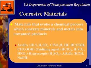
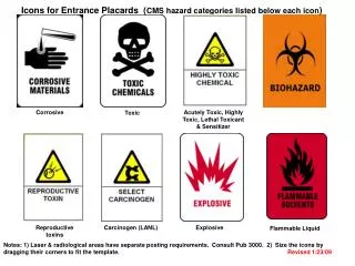
Substances • Alkalis: • Most cases of caustic injury in western countries • Cleaning agents (NaOH), drain openers, bleaches, toilet bowel cleaners, and detergents… • Acids • Less frequently in western countries; more common in countries like India (glacial acetic acid) • Toilet bowel cleaners ( sulfuric, hydrochloric ), anti rust compounds ( hydrochloric, oxalic, hydrofluoric ), swimming pool cleaners ( hydrofluoric )
Alkalis PH > 7 Tasteless, odorless →larger amounts liquefaction necrosis => direct extension, deeper injuries Solid form : limited quantities, oropharyngal and supraglottic injuries Liquid form: significant quantities, esophageal injury, extensive, circumferential burns Acid PH < 7 Pungent odor and noxious taste coagulation necrosis => formation of a coagulum layer : limit the depth of injury Less esophageal injury More gastric injury Alkalis V.S Acid
Pathophysiology • Alkali-induced injury: • Liquefactive necrosis • 1-2 days: Thrombosis of small vessels • 2-4 days : Newly forming blood vessels , fibroblasts migration • 4-7 days: Mucosal sloughing, bacterial invasion, inflammatory response, and development of granulation tissue • > 2 weeks: Collagen deposition • > 3 weeks: Scar retraction => may continue for several months • Acid-induced injury: • Superficial coagulation necrosis • Thromboses the underlying mucosal blood vessels and consolidates the connective tissue => Protective eschar
Pathologic severity of injury • First-degree: • Superficial mucosal damage • Focal or diffuse erythema, edema, hemorrhage • Without scar formation • Second-degree • Mucosal and sub-mucosal damage • Ulcerations, exudates, vesicle formation, granulation, fibroblastic reaction • Scar formation • Third-degree • Trans-mural • Deep ulcers and black discoloration and perforation of the wall
Clinical presentation • Vary widely • Hoarseness, stridor, dyspnea => Airway evaluation • Perforation: (During first 2 weeks) • Retro-sternal or back pain • Localized abdominal tenderness, rebound, rigidity, Psoas sign, obturator sign • Massive hematemesis • Dysphagia, odynophagia, drooling, nausea, vomiting • Early signs and symptoms may not correlate with the severity and extent of tissue injury • Oropharyngeal burns (-):10-30% esophageal burns(+) Oropharyngeal burns (+): 70% esophageal burns(-)
Diagnosis and staging • Upper gastrointestinal endoscopy • Endoscopic grading system • Grade 0: Normal • Grade 1: Mucosal edema and hyperemia • Grade 2A: Superficial ulcers, bleeding, exudates => Excellent prognosis • Grade 2B: Deep focal or circumferential ulcers • Grade 3A: Focal necrosis => Develop strictures: 70-100% • Grade 3B: Extensive necrosis => Early mortality rate: 65%
Late sequelae • Stricture formation • Primarily in those with grade 2B or 3 injury • Peak incidence: two months • Occur as early as two weeks or as late as years after ingestion • Gastric outlet obstruction • Early satiety , weight loss • Less frequently • 5-6 weeks ~ several years • Usually acid ingestion
Late sequelae • Esophageal carcinoma • Incidence: 1000 to 3000-fold increase • 3% have history of caustic ingestion • Mean latency: 41 years (13-71years) • Scar carcinoma: • Less distensible => dysphagia presents earlier • Lymphatic spread and direct extension • Surveillance • Begin 15-20 years after ingestion • The time interval : No more than every 1-3 years • Gastric carcinoma • rare occurrence
Management – General management • First aid • Identify the swallowed toxic agents • Avoid: • The use of emetics: re-exposes • Neutralizing agents: thermal injury • Gastric lavage: lead to perforation • Transfer to hospital immediately • Keep NPO • Insert NG tube ? • R/O perforation • Plain films of chest and abdomen • Esophagogram: Water-soluble agent • For ENT doctor • Airway evaluation • Oropharyngeal burns
Management - Endoscopy • Timing: • No later than 48 hours • Usually avoided from 5-15 days • Purpose: • Grading, manage appropriately • Risk of perforation: • Low, under adequate sedation • Extent: • Advance until a circumferential second-degree or third degree burn is seen • To first part of duodenum
Management - Oral intake • NPO before PES • Grade 1 or 2A injury: • A liquid diet may be initiated • Advance to a regular diet in 24 - 48 hours • Grade 2B or 3 injury: • Controversial • NG feeding, initiated after 24 hours => oral liquids are allowed after the first 48 hours if the patient is able to swallow saliva • TPN use with delayed oral feeding (7 days) => Avoid food irritation
Management - Prevention of strictures • Steroids • In animal studies: incidence of stricture formation • In human studies: Inconclusive so far • NEJM. 1990: • Prospective study over an 18-year period • No benefit • Related only to the severity of the corrosive injury • Toxicol Rev. 2005: • 1991-2004 in the English, German, French, Spanish • No benefit
Management - Prevention of strictures • Antibiotics • Decreased bacterial counts, reduction in inflammation • Mask the sign of more severe infection • A prophylactic antibiotic, in the absence of steroid therapy, is not advocated • Nasogastric tube • Feeding and stenting • Contribute to the development of long strictures • Routine use is not warranted
Management - Prevention of strictures • Total parental nutrition: • No human randomized study • NPO allowing the re-epithelialization • Intraluminal stent: • Controversial • Prevents opposite raw surfaces contact and decreases stricture formation (Gastrointest Endosc. 2004)
Management - Prevention of strictures • Early dilataion: • Less than one week • Controversial, most study: not recommended • Start during the 1st week => The stricture can resolve more easily (Pediatr Surg Int. 2005 ) • Anti-reflux treatment and Sucralfate: • Empirically use ; PPI, H2 blockers • Prevention of injured esophageal mucosa from gastric acid reflux
Management – Treatment of strictures • Endoscopic dilatation • The goal: dilate the esophageal lumen to 15 mm • Perforation rate: 0.5% • Special consideration: • Long, eccentric strictures: risk of perforation increased • Thick-walled strictures: recur rapidly • Multiple sessions: elective esophageal resection • Intraluminal stent • Temporary placement of a self-expanding plastic stent • Successful in case reports • Surgery • Esophagectomy with colonic interposition • Gastric transposition: high leak rate • Perform 6 months later
Conclusion • Signs and symptoms alone are an unreliable guide to injury • Early endoscopy has a crucial role • Grading, manage appropriately
Reference • Ramasamy K, Gumaste VV. Corrosive ingestion in adults. J Clin Gastroenterol. 2003;37:119-24. • Huang YC, Ni YH, Lai HS, Chang MH. Corrosive esophagitis in children.Pediatr Surg Int. 2004;20:207-10. • Pelclova D, Navratil T. Do corticosteroids prevent oesophageal stricture after corrosive ingestion? Toxicol Rev. 2005;24:125-9. • Baskin D, Urganci N, Abbasoglu L, et al. A standardised protocol for the acute management of corrosive ingestion in children.Pediatr Surg Int. 2004;20:824-8. • Anderson KD, Rouse TM, Randolph JG. A controlled trial of corticosteroids in children with corrosive injury of the esophagus. N Engl J Med. 1990;323:637-40. • Poley JW, Steyerberg EW, Kuipers EJ, et al. Ingestion of acid and alkaline agents: outcome and prognostic value of early upper endoscopy. Gastrointest Endosc. 2004;60:372-7. • Tiryaki T, Livanelioglu Z, Atayurt H. Early bougienage for relief of stricture formation following caustic esophageal burns. Pediatr Surg Int. 2005;21:78-80. • Evrard S, Le Moine O, Lazaraki G, et al. Self-expanding plastic stents for benign esophageal lesions.Gastrointest Endosc. 2004;60:894-900.