Download
1 / 15
150 likes | 182 Views
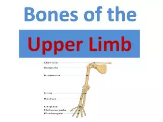
Discover the structure and functions of the pelvic girdle bones (Ilium, Ischium, Pubis), gender differences, and femur details. Learn about the tibia, fibula, and bones of the foot for a comprehensive understanding of the lower limb.

E N D
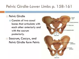
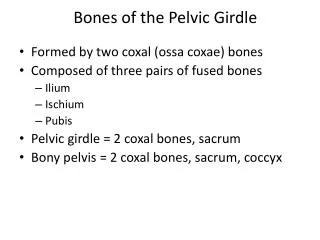
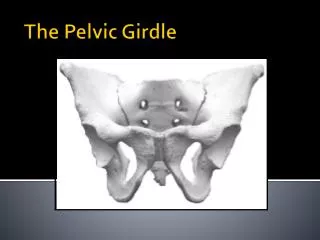
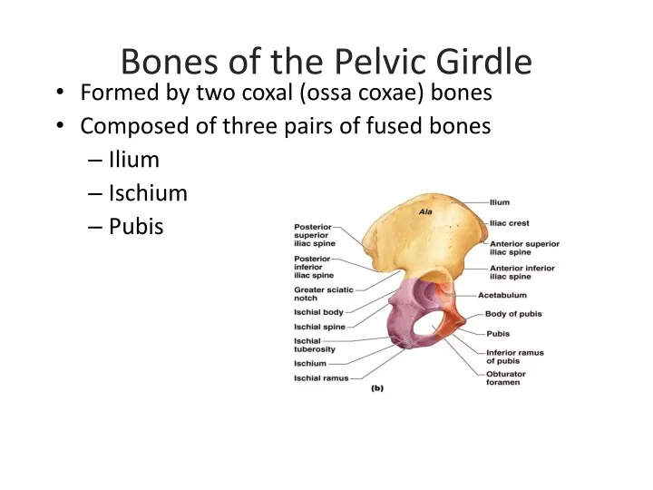
Bones of the Pelvic Girdle • Formed by two coxal (ossa coxae) bones • Composed of three pairs of fused bones • Ilium • Ischium • Pubis
Bones of the Pelvic Girdle • The total weight of the upper body rests on the pelvis • It protects several organs Reproductive organs Urinary bladder Part of the large intestine
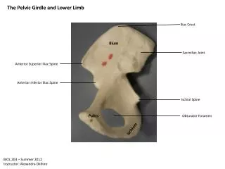
Ilium • Iliac crest – superior edge of “ala” • Greater Sciatic Notch – notch that allows blood vessels and the sciatic nerve to pass from the pelvis posteriorly into the thigh • Iliac Tuberosity - elevated and rough surface inferior to iliac crest, for the attachment of the posterior sacroiliac ligaments
Ilium • Iliac Fossa - large, smooth, concave surface on the internal surface of the • Acetabulum – Site where ilium, ischium, and pubis bones fuse. Deep socket that receives the head of the femur.
Ischium • Ischial Spine – superior to ischial tuberosity; projection that narrows pelvic outlet • Lesser Sciatic Notch – notch below ischial spine • Ischial Tuberosity – roughened area that receives body weight when sitting • Ischial Ramus – inferior portion of ischium that connects with pubis • Obturator Foramen – opening that allows blood vessels and nerves to pass into the anterior thigh
Pubis • Inferior Ramus – inferior portion that connects with ischium • Pubic tubercle - prominent forward-projecting structure on the upper border
Gender Differences of the Pelvis (check the book for a comparison photo) • The female inlet is larger and more circular • The female pelvis as a whole is shallower, and the bones are lighter and thinner • The female ilia flare more laterally • The female sacrum is shorter and less curved • The female ischial spines are shorter and farther apart; thus the outlet is larger • The female pubic arch is more rounded because the angle of the pubic arch is greater
Femur • Femur (thigh bone) • The heaviest, strongest bone in the body
Femur • Greater Trochanter – adjacent to head, most superior portion; site for muscle attachment • Lesser Trochanter – inferior to greater trochanter; site for muscle attachment • Head – round; articulates with acetabulum • Lateral Condyle – distal, lateral end of femur; articulates with tibia • Medial Condyle - distal, medial end of femur; articulates with tibia
Femur • Intercondylar fossa – separates lateral and medial condyles on posterior side of femur • Popliteal surface - triangular area (“back of knee”) between medial and lateral condyles. Patella = knee cap
Tibia and Fibula • Tibia • Shinbone • Larger and medially oriented • Fibula • Thin and sticklike
Tibia • Lateral condyle - at proximal end; articulates with lateral condyle of femur to create knee joint • Medial condyle – at proximal end; articulates with medial condyle of femur to create knee joint • Tibial tuberosity – roughened area on anterior surface where patellar tendon attaches • Medial malleolus – distal end, process that creates inner bulge of ankle
Fibula • Head – proximal end; articulates with inferior portion of lateral condyle of tibia (not part of knee joint) • Lateral malleolus – distal end; forms outer part of the ankle
Tarsals, Metatarsals,Phalanges • Tarsals • Two largest tarsals • Calcaneus (heelbone) • Talus • Metatarsals—sole • Phalanges—toes