Download
1 / 19
200 likes | 258 Views
Explore how neurotransmitters affect neuron communication, from synaptic cleft to receptor binding. Learn about excitatory and inhibitory synapses, EPSPs, IPSPs, and neurotransmitter classifications. Discover how neural integration occurs in neuronal pools through various circuits.

E N D
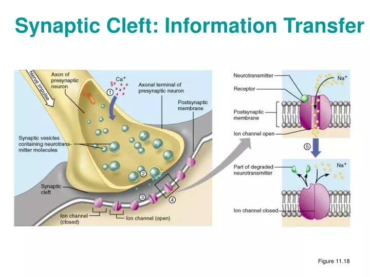
Synaptic Cleft: Information Transfer Figure 11.18
Termination of Neurotransmitter Effects • Neurotransmitter bound to a postsynaptic neuron: • Produces a continuous postsynaptic effect • Blocks reception of additional “messages” • Must be removed from its receptor • Removal of neurotransmitters occurs when they: • Are degraded by enzymes • Are reabsorbed by astrocytes or the presynaptic terminals • Diffuse from the synaptic cleft
Synaptic Delay • Neurotransmitter must be released, diffuse across the synapse, and bind to receptor • Synaptic delay – time needed to do this (0.3-5.0 ms) • Synaptic delay is the rate-limiting step of neural transmission
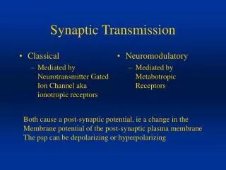
Postsynaptic Potentials • Neurotransmitter receptors mediate changes in membrane potential according to: • The amount of neurotransmitter released • The amount of time the neurotransmitter is bound to receptor • The two types of postsynaptic potentials are: • EPSP – excitatory postsynaptic potentials • IPSP – inhibitory postsynaptic potentials
Excitatory Postsynaptic Potentials • EPSPs are graded potentials that can initiate an action potential in an axon • Use only chemically gated channels • Na+ and K+ flow in opposite directions at the same time • Postsynaptic membranes do not generate action potentials Figure 11.19a
Inhibitory Synapses and IPSPs • Neurotransmitter binding to a receptor at inhibitory synapses: • Causes the membrane to become more permeable to potassium and chloride ions • Leaves the charge on the inner surface negative • Reduces the postsynaptic neuron’s ability to produce an action potential Figure 11.19b
Summation • A single EPSP cannot induce an action potential • EPSPs must summate temporally or spatially to induce an action potential • Temporal summation – presynaptic neurons transmit impulses in rapid-fire order • Spatial summation – postsynaptic neuron is stimulated by a large number of terminals at the same time • IPSPs can also summate with EPSPs, canceling each other out
Summation Figure 11.20
Neurotransmitters • Chemicals used for neuronal communication with the body and the brain • 50 different neurotransmitter have been identified • Classified chemically and functionally
Chemical NeurotransmittersLEARN CHART • Acetylcholine (ACh) • Biogenic amines • Amino acids • Peptides • Novel messengers
Functional Classification of Neurotransmitters • Two classifications: excitatory and inhibitory • Excitatory neurotransmitters cause depolarizations (e.g., glutamate) • Inhibitory neurotransmitters cause hyperpolarizations (e.g., GABA and glycine) • Some neurotransmitters have both excitatory and inhibitory effects • Determined by the receptor type of the postsynaptic neuron • Example: aceytylcholine • Excitatory at neuromuscular junctions • Inhibitory with cardiac muscle
Neurotransmitter Receptor Mechanisms • Direct: neurotransmitters that open ion channels • Promote rapid responses • Examples: ACh and amino acids • Indirect: neurotransmitters that act through second messengers • Promote long-lasting effects • Examples: biogenic amines and peptides
ANIMATIONS • Salutatory conduction • http://www.blackwellscience.com/matthews/actionp.html • terminal end • http://www.mind.ilstu.edu/flash/synapse_1.swf • Inhibitory • http://www.blackwellscience.com/matthews/neurotrans.html • Paxil and serotonin • http://www.paxil.com/flash/depression.swf
Cocaine and Crack • Blocks reuptake of Dopamine • Great at first, but not after Dopamine is degraded • Moods
G Protein-Linked Receptors: Mechanism • Neurotransmitter binds to G protein-linked receptor • G protein is activated and GTP is hydrolyzed to GDP • The activated G protein complex activates adenylate cyclase • Adenylate cyclase catalyzes the formation of cAMP from ATP • cAMP, a second messenger, brings about various cellular responses
Neural Integration: Neuronal Pools • Functional groups of neurons that: • Integrate incoming information • Forward the processed information to its appropriate destination
Neural Integration: Neuronal Pools • Simple neuronal pool • Input fiber – presynaptic fiber • Discharge zone – neurons most closely associated with the incoming fiber • Facilitated zone – neurons farther away from incoming fiber Figure 11.23
Types of Circuits in Neuronal Pools • Divergent – one incoming fiber stimulates ever increasing number of fibers, often amplifying circuits Figure 11.24a, b