Download
1 / 63
730 likes | 1.9k Views
Fractures of the Tibia and Fibula in the Pediatric Patient. Steven Rabin MD Revised February 2011 First Edition Created by Steven Frick , MD Created March 2004; Revised August 2006. Growth and Development of the Tibia and Fibula. Most growth from proximal physis

E N D
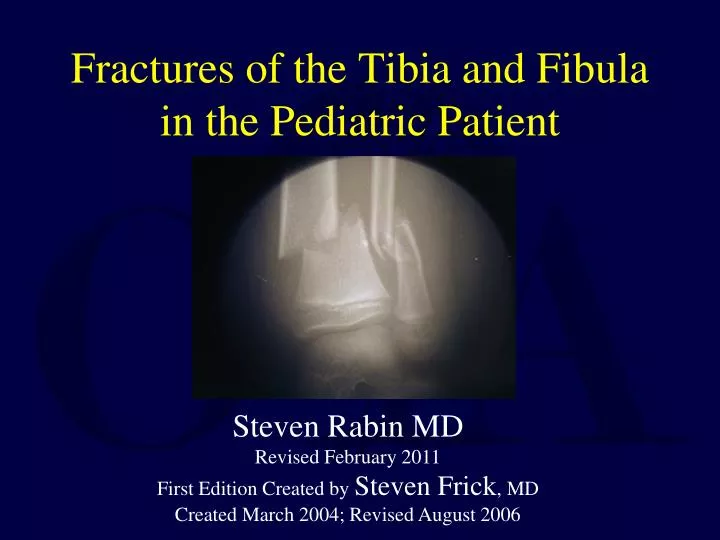
Fractures of the Tibia and Fibula in the Pediatric Patient Steven Rabin MD Revised February 2011 First Edition Created by Steven Frick, MD Created March 2004; Revised August 2006
Growth and Development of the Tibia and Fibula • Most growth from proximal physis • Fibula moves posterior to the tibia with growth • Extra-physeal fractures rarely disturb future growth and development • Exceptions= proximal tibia, cross-union
Relevant Anatomy • Tibia and fibula bound together by interosseous membrane • Some motion occurs normally (proximal distal translation, inward/outward rotation) at proximal and distal tibia-fibular joints • Subcutaneous location of tibia- implications for susceptibility to injury and healing potential
Incidence • Low energy fractures common (toddler’s fracture, spiral tibia fractures) • Tibia is the most frequent location for open fractures in children
History • As with all Pediatric Fractures: • A high index of suspicion for Child Abuse should be maintained. • Especially with a history of multiple long bone fractures, fractures in non-ambulatory patients, and history of multiple fractures • Be alert for other causes of multiple fractures such as osteogenesis imperfecta.
Physical Examination • Integrity of the skin and severity of soft tissue injury • Dorsalis pedis and posterior tibialis pulsers, distal capillary refill • Peroneal and posterior tibial nerve function • Any signs of compartment syndrome
Radiographic Evaluation • Two orthogonal views usually adequate • Visualize knee and ankle joints • Assess for displacement, translation, shortening, angulation • Rotation best assessed clinically
Classification • Open/Closed • Tibia, Fibula or both • Fracture location- proximal or distal, metaphyseal or diaphyseal • Fracture pattern- transverse, spiral, butterfly, comminuted • Involvement of Growth Plates
Decision Making-Principles for Treatment • Restore acceptable length, alignment, translation and rotation • What is acceptable? Little hard data available, depends on age of patient • General guidelines for acceptable position: <10° angulation, <2cm shortening, <50% translation, rotation equal to opposite side
Remodeling – General Guidelines • Fractures that heal in positions outside these guidelines may remodel or go on to good clinical result – BUT May Not! • Children <10 years old have more potential to remodel • Remodeling more reliable in plane of joint motion • Metaphyseal fractures remodel better than diaphyseal • Do not expect rotational deformity to remodel • Overgrowth can occur but not predictable
Principles of Treatment • Majority of tibia/fibula fractures in children can be treated closed • Above knee vs. below knee cast • Radiographic monitoring at regular intervals during early healing- wedge cast, or remanipulate/recast for unacceptable reduction/position
Treatment Options • Cast above knee usually, but below knee acceptable for stable fracture patterns or after early healing • Pin fixation and cast- simple and effective, especially in oblique fractures, younger children • External fixation- high energy fractures with associated soft tissue injuries • Flexible nails- proximal medial and lateral insertion • Rigid nail- if near skeletal maturity • Plate fixation- if soft tissues allow
Specific Fractures • Proximal Tibia Physis Injury • Tibial Tuberosity Avulsion • Toddlers • Proximal tibia metaphyseal fracture • Isolated fibula fracture • Isolated tibia fracture- mid/distal third • Open tibia fracture • Distal metaphyseal tibia fracture • Floating knee • Pathologic Fracture
PROXIMAL TIBIA GROWTH PLATE INJURIES • <1% of all physeal injuries: proximal growth plate is protected by ligamentous attachments and fibula • Proximal Physis • .65cm growth/year • 45% length of tibia, 27% length of leg • Most common injury is 11-14y/o boys with hyperextension • Neurovascular compromise possible with posterior displacement • Treatment: if nondisplaced: cast 3 -4 weeks. If displaced, closed reduction/above knee cast for 4 weeks. No sports for 4 months.
Tibial Tuberosity Apophysis Avulsion Fractures • Mechanism: Jump or landing where Quadriceps contraction pulls off the tubercle as the knee flexes. • Growth disturbance rare but • Genu recurvatum possible especially in patients less than 11 y/o
Tibial Tuberosity: Treatment • Nondisplaced fractures: cast for 6 weeks with the knee extended. • Displaced fractures require open reduction with internal fixation
Tibial Tuberosity Apophysis • ORIF
Toddler’s Fracture • Isolated tibia fracture • Very common in young children • Usually twisting injury • Stable injuries • Treatment: • If distal, short leg cast for 3-4 weeks • If proximal, above knee cast with knee flexed 10 degrees for 3-4 weeks, but many treated with immediate short leg cast
Proximal Tibia Metaphyseal Fracture • Usually 3-6 years old when femoral- tibial angle is growing towards valgus • Tendency toward valgus overgrowth • Varus mold may prevent • Valgus can be severe but usually remodels over years such that corrective osteotomy unnecessary
Valgus after Proximal Tibia Metaphyseal Fracture Asymmetric growth slowdown lines Persistent bow
Valgus following Proximal Tibia Fracture Case courtesy of K. Shea. Observe and often improve with time, but may need guided growth surgical intervention
Isolated Fibula Fractures • Direct blow mechanism • Immobilize as needed for comfort(fibula 15% of weight bearing) • Carefully assess ankle (r/o Maissenouve injury)
Tibia Shaft Fractures • 5% of all pediatric fractures • 70% have intact fibula, 30% both bones fractured
Isolated Tibia Fracture with Intact Fibula • Often at middle/distal third • Muscle forces/biomechanics usually result in drift into varus angulation • Valgus mold in initial cast • Can wedge at 2 weeks but more difficult because of intact fibula
Example:Isolated Tibia Fracture • Isolated Tibia Fracture Casted with Valgus Mold – Healed with Excellent Alignment
Below knee cast may be adequate for all pedi tibia fxs Klatt, et al. OTA 2010 • Retrospective cohort study • 269 pediatric tibial shaft fractures without fibula shaft fracture • No sig. malunion or loss of alignment with below knee cast treatment compared to above knee cast
Indications for Surgical Treatment • Inability to obtain/maintain acceptable position • Open fractures • Multiple trauma to facilitate mobilization
Open Tibia Fractures • Soft tissue injuries typically less severe than in adults • Periosteum often intact on concavity • Appropriate timely debridement, antibiotics • Pins and cast, external fixation, flexible intramedullary rods all useful – choice depends on age, fracture pattern, status of soft tissues, associated injuries • Lower malunion rates and best outcomes seem to be reported after flexible nailing
Open Tibia Fracture with Soft Tissue Deficits Appropriate pin placement and construct needed to control varus
Distal Metaphyseal Tibia Fracture • “Gillespie” fracture – apex posterior angulation of the distal tibia • Dorsiflexion of ankle to neutral may increase apex posterior angulation • Cast in equinus until early healing, then change cast and dorsiflex to neutral
Gillespie Fracture – Healed in Excessive DF as was Casted with Ankle at Neutral
Pinning and Cast after Failure to Achieve Acceptable Alignment with Closed Methods
Expected Outcomes for Tibia Fractures • Heal in 6-12 weeks in juveniles/adolescents • Heal in 3-4 weeks in toddlers • Nonunions are rare
Complications • Compartment syndrome • Malunion • Growth arrest/deformity
Compartment Syndrome • Can occur in skeletally immature patient after closed or open tibia fracture • Reported in up to 5-10% of Pediatric Tibia Shaft fractures • Tense compartment, pain out of proportion, pain with passive stretch, paresthesias in distribution of nerves that are in compartment • Compartment pressure measurement to confirm • Consider conscious sedation / general anesthesia in child to measure pressures • Fasciotomies emergently if diagnosed • COMPARTMENT SYNDROME CAN OCCUR EVEN IN APPARENT “LOW ENERGY” SPIRAL PATTERNS
Malunions • No Consistent Literature to define Malunion • Long term adverse effects of Malunion not well documented. • General guidelines for acceptable position: <10° angulation, <2cm shortening, <50% translation, rotation equal to opposite side
Tibia Fracture Malunion/Nonunion • Typical Malunion Deformity is Varus
Malunion Example • Varus – procurvatum malunion following premature removal of external fixator after open tibia fracture
Malunion Example • Treatment with Osteotomy and Plate Fixation