Download
1 / 77
800 likes | 1.17k Views
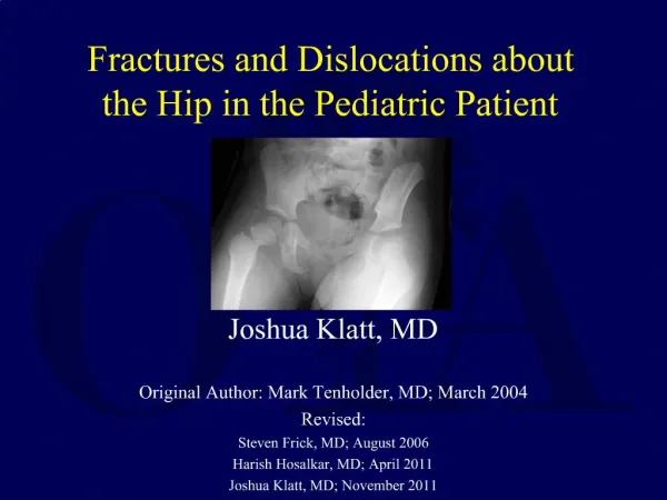
Fractures and Dislocations about the Hip in the Pediatric Patient. Joshua Klatt, MD Original Author: Mark Tenholder, MD; March 2004 Revised: Steven Frick, MD; August 2006 Harish Hosalkar, MD; April 2011 Joshua Klatt, MD; November 2011.

E N D
Fractures and Dislocations about the Hip in the Pediatric Patient Joshua Klatt, MD Original Author: Mark Tenholder, MD; March 2004 Revised: Steven Frick, MD; August 2006 Harish Hosalkar, MD; April 2011 Joshua Klatt, MD; November 2011
“Hip fractures in children are of interest because of the frequency of complications rather than the frequency of fractures.” • Canale
Femoral Neck Fractures in Children • Rare fracture • Anatomic and vascular differences • Emergent treatment • High complication rate
Background • Different from Adults • High-energy • Thick periosteum • Vascularity • Physes • Treatment options
Background • Osseous Anatomy • Proximal femoral physis • Trochanteric apophysis • Dense bone • Small neck
Background • Vascular Anatomy • Immature • Variable • Ligamentum teres • Lateral epiphyseal vessels (bypass physis) • Metaphyseal circulation (after physeal closure) • Vulnerable to injury
Mechanism • MVC • Auto-ped • High falls • Minor trauma can still be a cause
Classification • Type 1 – Transepiphyseal • Type 2 – Transcervical • Type 3 – Cervicotrochanteric • Type 4 - Intertrochanteric Colonna PC. Fractures of the neck of the femur in children. Am J Surg 1929;6:793-7.
Type I • Very rare • Little evidence • High risk of AVN (up to 100% in some series) • Canale ST, Bourland WL. Fracture of the neck and intertrochanteric region of the femur in children. J Bone Joint Surg Am. 1977 Jun.;59(4):431–443.
Type ITreatment • Nondisplaced • Can treat with spica cast • Displaced • Past • Closed reduction and spica • ORIF • Present • Closed or open reduction plus internal fixation • Threaded pins • Cannulated screws • Smooth pins • Forlin E, Guille JT, Kumar SJ, Rhee KJ. Transepiphyseal fractures of the neck of the femur in very young children. J Pediatr Orthop. 1992 Feb.;12(2):164–168.
Type IResults • Recent literature following better understanding of hip vascularity • In some circumstances the femoral head may not be completely avascular, and, with appropriate surgical care, the hip can be preserved Schoenecker JG, Kim Y-J, Ganz R. Treatment of traumatic separation of the proximal femoral epiphysis without development of osteonecrosis: a report of two cases. The Journal of Bone and Joint Surgery. 2010 Apr.;92(4):973–977.
Type IExample • 10 yr female • Type I fracture-dislocation of hip
Type IExample ORIF and Pins Attempted
Type IExample • Postop film • Malreduced and dislocated
Type IExample Repeat ORIF
Type IExample 3 month follow-up
Type IExample 8 Months Heterotopic ossification evident
Type IExample • 11 Months • Osteonecrosis
Type II • Most common type (50% of peds hip fx) • Most common AVN (50%) • 3/4 will be displaced
Type II Historical treatment Internal fixation is currently the treatment of choice • Lam. Fractures of the neck of the femur in children. J Bone Joint Surg Am. 1971;53:1165–1179. • Ratliff. Fractures of the neck of the femur in children. J Bone Joint Surg Br. 1962;44-B:528–542. • Canale ST, Bourland WL. Fracture of the neck and intertrochanteric region of the femur in children. J Bone Joint Surg Am. 1977;59:431–443. • Quick. Pediatric Fractures and Dislocations of the Hip and Pelvis. Clin Orthop Relat Res. 2005;(432):87–96.
Type IITreatment • Nondisplaced • Spica cast, if young • Use internal fixation, if older • If in doubt, treat as displaced
Type IITreatment • Displaced • Anatomic reduction is important, open if necessary • Do not accept varus mal-reductions • Avoid excess traction • Fracture table may be used without extreme positioning for prolonged period • Cannulated screws/ threaded pins to compress • Avoid physis • But stability and reduction is first priority
Type IIResults • Nondisplaced • Fewer complications • Outcome in literature is variable • AVN in up to 50% • Highest complication rate of the 4 types • Improved with internal fixation İnan U, Köse N, Ömeroğlu H. Pediatric femur neck fractures: a retrospective analysis of 39 hips. J Child Orthop. 2009 May 26;3(4):259–264.
Type III • Second most common • 35% of peds hip fx • Second highest AVN rate • 25-30% • 2/3 displaced İnan U, Köse N, Ömeroğlu H. Pediatric femur neck fractures: a retrospective analysis of 39 hips. J Child Orthop. 2009 May 26;3(4):259–264.
Type IIITreatment • Nondisplaced • Spica cast • Follow closely for loss of reduction • Displaced • ORIF • Cannulated screws • Peds hip screw • Avoid physes İnan U, Köse N, Ömeroğlu H. Pediatric femur neck fractures: a retrospective analysis of 39 hips. J Child Orthop. 2009 May 26;3(4):259–264.
Type IIIResults • Slightly better than II • Nondisplaced • Fewer complications • Outcome in literature is variable • AVN in up to 30% • IF reduces coxa vara and nonunion Flynn. Displaced fractures of the hip in children. Management by early operation and immobilisation in a hip spica cast. J Bone Joint Surg Br. 2002;84:108–112.
Type IIIExample • 6 year old femal • MVC • Liver laceration • Ipsilateral femoral neck, femur, and tibia fractures
Type IIIExample • 8 wks post-op • Union • Cast removed, WBAT • No AVN
Type IV • Not common • 10-15% of peds hip fx • Fewest complications • AVN still possible, but unusual
Type IVTreatment • Most agreement between authors • Nondisplaced • Hip-spica in younger patients • Displaced • Pediatric hip screw in older pts • Or in those with unstable reduction
Type IVResults • Generally good • Fewest complications • High energy still can result in AVN (10-20%)
Type IVExample 14 year old male Motorcycle crash
Type IVExample 9 weeks post-op
Type IVExample 9 months post-op
Type IVExample 10 months post-op After hardware removal
Type IVExample 15 months post-op AVN
Hip FractureTreatment Highlights • Data on nondisplaced fractures is limited • Conclusions are difficult • Most nondisplaced fractures can be treated in a cast • Exceptions • Older child • Type II
Hip FractureTreatment Highlights • Surgery and implants available now are different than those used in older literature • More recent emphasis on internal fixation • Anatomic reduction and compression is key for successful union • Surgical approach should not further destabilize blood supply to femoral head • Expanded indications in polytrauma pt’s
Hip FractureComplications • Lam. Fractures of the neck of the femur in children. J Bone Joint Surg Am. 1971;53:1165–1179. • Ratliff. Fractures of the neck of the femur in children. J Bone Joint Surg Br. 1962;44-B:528–542. • Canale ST, Bourland WL. Fracture of the neck and intertrochanteric region of the femur in children. J Bone Joint Surg Am. 1977;59:431–443.
Hip FractureAVN Most common and devastating complication
Hip FractureAVN • 6 – 53% overall rate • Type I 57% to 100% • Type II 50% • Type III 25% • Type IV 10% Quick TJ, Eastwood DM. Pediatric Fractures and Dislocations of the Hip and Pelvis. Clin Orthop Relat Res. 2005;432:87–96.
Hip FractureAVN • AVN may develop if • The vessels are torn in the initial injury • The vessels are kinked at due to displacement • There is intracapsular tamponade causing vascular disruption • The vessels are not protected during healing Quick TJ, Eastwood DM. Pediatric Fractures and Dislocations of the Hip and Pelvis. Clin Orthop Relat Res. 2005;432:87–96.
Hip FractureAVN • Factors influencing rate of AVN • Degree of initial displacement • Timing of reduction and fixation • Quality of reduction • Stability of reduction and fixation • Decompression of capsular hematoma • Weight-bearing status Quick TJ, Eastwood DM. Pediatric Fractures and Dislocations of the Hip and Pelvis. Clin Orthop Relat Res. 2005;432:87–96.
AVNClassification Ratliff 1962 Ratliff A. Fractures of the neck of the femur in children. J Bone Joint Surg Br. 1962 Aug.;44-B:528–542.