Download
1 / 42
430 likes | 636 Views
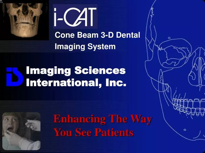
Imaging Sciences International, Inc. Cone Beam 3-D Dental Imaging System. Enhancing The Way You See Patients. Who is Imaging Sciences International?. Meeting the Advanced Imaging Needs of Dental Professionals Since 1992.

E N D
Imaging Sciences International, Inc. Cone Beam 3-D Dental Imaging System Enhancing The Way You See Patients
Who is Imaging Sciences International? Meeting the Advanced Imaging Needs of Dental Professionals Since 1992 • Since 1992, Imaging Sciences International, Inc. has been a global leader in developing the most advanced computer controlled dental and maxillofacial radiography products. • ISI’s marquee products: i-CAT Panorex CMT CommCAT • Imaging Sciences designs, develops, and produces cutting-edge imaging technologies to provide superior quality and function that enhance the way doctors see patients. ISI is dedicated to continually advancing imaging innovation in the dental industry, as well as outstanding customer service.
Why Cone Beam 3-D Dental Imaging? Societal Drivers • Patients are increasingly demanding real-time treatment options (instant crowns, veneers, over-dentures, implants, and braces) • Increased demand for cosmetic dentistry and willingness to invest in elective (out-of-pocket) procedures Technology Drivers • Digital – paperless office • True anatomical information - Distortion-Free • DICOM 3 information for pre-surgical 3-D planning software, SLA models, and drill guides Benefits to the Dental Practice • Increase productivity- generate more profit per unit chair time • Increase case acceptance- uncompleted treatment proposals are the largest non value-added activity for dentists • Surgical predictability • A complete continuum of care- diagnosis through treatment
Why Cone Beam 3-D Dental Imaging? Cone Beam 3-D images provide high-definition, three dimensional, digital data on precise anatomical information of all oral and maxillofacial structures ~At reduced cost to the doctor ~With less radiation to the patient Vs. Traditional imaging systems, which are limited by distortion, magnification changes, restricted clarity, lack of accuracy in measurements, and not allowing for 3-D modeling
Why Enhancing The Way You See Patients • Surgical Predictability • Saves Surgery Time/Reduce Costs • Keeps Patients In-House • Close More Cases/Increase Revenue • Decreased Medical & Legal Risks
Cone Beam 3-D Imaging System • 3-D volumetric images • True anatomic measurements • 14 BIT Gray Scale • Pan-sized footprint • Fast Scan Time • Low Radiation • Higher resolution for all views (amorphous silicon flat panel image sensor)
Effective Dose Comparison • i-CAT 20 second scan: 68 uSv Exposure is in “Pulsed” mode, actual exposure time is about 3.5 seconds for a 20 second scan • i-CAT 10 second scan: 34 uSv • Daily background: 8 uSv • Panoramic (Average): 10-15 uSv Digital Panoramic 4.7 – 14.9 uSv Highest Film Pan 26 uSv • Full mouth series: 150 uSv • Medical CT 1200-3300 uSv* Dr. Sharon Brooks, Dept. of Radiology, University of Michigan *Dr. Stuart White, Dept. of Radiology, UCLA
Cone Beam 3-D Imaging System Enhancing The Way You See Patients • Implant Studies • Impaction • Orthognathic Surgery • TMJ Analysis • Airway Studies • Spinal Studies • Orthodontics
for Implants Provides Complete 3-D Information to optimize treatment planning and placement • Locate critical anatomy • Determine if bone grafting or sinus lift is warranted • Select the most suitable implant size and type • Optimize locations and angulations • Coordinate with Restorative Dentist
3-D Implant Visualization and Planning DICOM 3 3rd party software. Please call for details.
for Implant 3-D Modeling and Drill Guides DICOM 3 3rd party software. Please call for details.
for Impaction More accurate 3-D views of impacted teeth • Provides more accurate 3-D views of impacted molars, impacted cuspids, and other supernumerary anomalies • Visualize impaction within the alveolar bone, location relative to adjacent teeth, and proximity to vital structures • More accurate information can result in less invasive surgery/decreased surgical time
Reposition for Orthognathic Surgery DICOM 3 3rd party software. Please call for details
for TMJ Analysis 3-D views of critical structures for complete TMJ Analysis • Provide Distortion-Free 3-D views of critical anatomy surrounding the condyles • Diagnosis of bone morphology, joint space, and function • High speed-scan for open jaw views • All done quickly and comfortably
for Airway Studies 3-D data enhances airway assessment • Assess airways and determine appropriate treatments • Identify restricted airways, which may be susceptible to collapse • Diagnose infected sinuses
for Orthodontics Improving Orthodontic diagnosis and treatment • Provide multiple projection perspective • Locate and diagnose pathologies • View impacted or supernumerary teeth • Assess tooth relationship to other teeth and anatomical structures
for Orthodontics Mandible Crowns Maxillary Crowns Occlusion Midsagittal Slice Coronal First Molar Region
for Orthodontics MIP View (Maximum Intensity Projection)
for Orthodontics 3-DVR™ (3-D Volume Rendering Software)
for Orthodontics 3-DVR™ (3-D Volume Rendering Software)
for Orthodontics DICOM 3 3rd party software. Please call for details
for Orthodontics DICOM 3 3rd party software. Please call for details
for Orthodontics DICOM 3 3rd party software. Please call for details
for Orthodontics DICOM 3 3rd party software. Please call for details
for Time Reduces: • Time and complication from surprises • Time for developing and duplicating x-rays • Time for retrieving x-rays from charts • Time for moving x-rays from operatory to operatory
for Referrals Internal Marketing: • Patients are amazed with the technology • Patients relate high technology with high quality • Patients leave with i-CAT reports reminding them of their experience with advanced technology and low radiation • The report print-outs contain the Dentists’ Name, Address, and Phone Number • Patients talk about your technology with employers, friends, and family
for Recognition Designates Recognition: • Leader in the Field • Quality Dental Care • High Technology Office • Caring Dentist
Why Enhancing The Way You See Patients • Surgical Predictability • Saves Surgery Time/Reduce Costs • Keeps Patients In-House • Close More Cases/Increase Revenue • Decreased Medical & Legal Risks
Imaging Sciences International 1-800-205-3570 www.ImagingSciences.com Info@ImagingSciences.com Enhancing The Way You See Patients