Download
1 / 123
1.23k likes | 1.26k Views
Explore the intricate structures of the eye, from eyelids to optic nerves, and learn about how each component contributes to vision. Discover the importance of sensory receptors, accessory structures, and the different tunics that make up the eyeball.

E N D
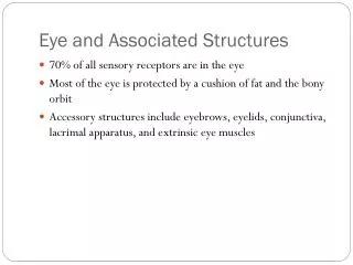
Eye and Associated Structures • 70% of all sensory receptors are in the eye • Most of the eye is protected by a cushion of fat and the bony orbit • Accessory structures include eyebrows, eyelids, conjunctiva, lacrimal apparatus, and extrinsic eye muscles
Eyebrows • Coarse hairs that overlie the supraorbital margins • Functions include: • Shading the eye • Preventing perspiration from reaching the eye • Orbicularis muscle – depresses the eyebrows • Corrugator muscles – move the eyebrows medially
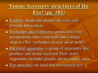
Palpebrae (Eyelids) • Protect the eye anteriorly • Palpebral fissure – separates eyelids • Canthi – medial and lateral angles (commissures)
Palpebrae (Eyelids) • Lacrimal caruncle – contains glands that secrete a whitish, oily secretion (Sandman’s eye sand) • Tarsal plates of connective tissue support the eyelids internally • Levator palpebrae superioris – gives the upper eyelid mobility
Palpebrae (Eyelids) • Eyelashes • Project from the free margin of each eyelid • Initiate reflex blinking • Lubricating glands associated with the eyelids • Meibomian glands and sebaceous glands • Ciliary glands lie between the hair follicles
Palpebrae (Eyelids) Figure 15.1b
Conjunctiva • Transparent membrane that: • Lines the eyelids as the palpebral conjunctiva • Covers the whites of the eyes as the ocular conjunctiva • Lubricates and protects the eye
Lacrimal Apparatus • Consists of the lacrimal gland and associated ducts • Lacrimal glands secrete tears • Tears • Contain mucus, antibodies, and lysozyme • Enter the eye via superolateral excretory ducts • Exit the eye medially via the lacrimal punctum • Drain into the nasolacrimal duct
Lacrimal Apparatus Figure 15.2
Extrinsic Eye Muscles • Six straplike extrinsic eye muscles • Enable the eye to follow moving objects • Maintain the shape of the eyeball • Four rectus muscles originate from the annular ring • Two oblique muscles move the eye in the vertical plane
Extrinsic Eye Muscles Figure 15.3a, b
Summary of Cranial Nerves and Muscle Actions • Names, actions, and cranial nerve innervation of the extrinsic eye muscles Figure 15.3c
Structure of the Eyeball • A slightly irregular hollow sphere with anterior and posterior poles • The wall is composed of three tunics – fibrous, vascular, and sensory • The internal cavity is filled with fluids called humors • The lens separates the internal cavity into anterior and posterior segments
Structure of the Eyeball Figure 15.4a
Fibrous Tunic • Forms the outermost coat of the eye and is composed of: • Opaque sclera (posteriorly) • Clear cornea (anteriorly) • The sclera protects the eye and anchors extrinsic muscles • The cornea lets light enter the eye
Vascular Tunic (Uvea): Choroid Region • Has three regions: choroid, ciliary body, and iris • Choroid region • A dark brown membrane that forms the posterior portion of the uvea • Supplies blood to all eye tunics
Vascular Tunic: Ciliary Body • A thickened ring of tissue surrounding the lens • Composed of smooth muscle bundles (ciliary muscles) • Anchors the suspensory ligament that holds the lens in place
Vascular Tunic: Iris • The colored part of the eye • Pupil – central opening of the iris • Regulates the amount of light entering the eye during: • Close vision and bright light – pupils constrict • Distant vision and dim light – pupils dilate • Changes in emotional state – pupils dilate when the subject matter is appealing or requires problem-solving skills
Pupil Dilation and Constriction Figure 15.5
Sensory Tunic: Retina • A delicate two-layered membrane • Pigmented layer – the outer layer that absorbs light and prevents its scattering • Neural layer, which contains: • Photoreceptors that transduce light energy • Bipolar cells and ganglion cells • Amacrine and horizontal cells
Sensory Tunic: Retina Figure 15.6a
The Retina: Ganglion Cells and the Optic Disc • Ganglion cell axons: • Run along the inner surface of the retina • Leave the eye as the optic nerve • The optic disc: • Is the site where the optic nerve leaves the eye • Lacks photoreceptors (the blind spot)
The Retina: Ganglion Cells and the Optic Disc Figure 15.6b
The Retina: Photoreceptors • Rods: • Respond to dim light • Are used for peripheral vision • Cones: • Respond to bright light • Have high-acuity color vision • Are found in the macula lutea • Are concentrated in the fovea centralis
Blood Supply to the Retina • The neural retina receives its blood supply from two sources • The outer third receives its blood from the choroid • The inner two-thirds is served by the central artery and vein • Small vessels radiate out from the optic disc and can be seen with an ophthalmoscope
Inner Chambers and Fluids • The lens separates the internal eye into anterior and posterior segments • The posterior segment is filled with a clear gel called vitreous humor that: • Transmits light • Supports the posterior surface of the lens • Holds the neural retina firmly against the pigmented layer • Contributes to intraocular pressure
Anterior Segment • Composed of two chambers • Anterior – between the cornea and the iris • Posterior – between the iris and the lens • Aqueous humor • A plasmalike fluid that fills the anterior segment • Drains via the canal of Schlemm • Supports, nourishes, and removes wastes
Anterior Segment Figure 15.8
Lens • A biconvex, transparent, flexible, avascular structure that: • Allows precise focusing of light onto the retina • Is composed of epithelium and lens fibers • Lens epithelium – anterior cells that differentiate into lens fibers • Lens fibers – cells filled with the transparent protein crystallin • With age, the lens becomes more compact and dense and loses its elasticity
Light • Electromagnetic radiation – all energy waves from short gamma rays to long radio waves • Our eyes respond to a small portion of this spectrum called the visible spectrum • Different cones in the retina respond to different wavelengths of the visible spectrum
Light Figure 15.10
Refraction and Lenses • When light passes from one transparent medium to another its speed changes and it refracts (bends) • Light passing through a convex lens (as in the eye) is bent so that the rays converge to a focal point • When a convex lens forms an image, the image is upside down and reversed right to left
Refraction and Lenses Figure 15.12a, b
Focusing Light on the Retina • Pathway of light entering the eye: cornea, aqueous humor, lens, vitreous humor, and the neural layer of the retina to the photoreceptors • Light is refracted: • At the cornea • Entering the lens • Leaving the lens • The lens curvature and shape allow for fine focusing of an image
Focusing for Distant Vision • Light from a distance needs little adjustment for proper focusing • Far point of vision – the distance beyond which the lens does not need to change shape to focus (20 ft.) Figure 15.13a
Focusing for Close Vision • Close vision requires: • Accommodation – changing the lens shape by ciliary muscles to increase refractory power • Constriction – the pupillary reflex constricts the pupils to prevent divergent light rays from entering the eye • Convergence – medial rotation of the eyeballs toward the object being viewed
Focusing for Close Vision Figure 15.13b
Problems of Refraction • Emmetropic eye – normal eye with light focused properly • Myopic eye (nearsighted) – the focal point is in front of the retina • Corrected with a concave lens • Hyperopic eye (farsighted) – the focal point is behind the retina • Corrected with a convex lens
Problems of Refraction Figure 15.14a, b
Photoreception: Functional Anatomy of Photoreceptors • Photoreception – process by which the eye detects light energy • Rods and cones contain visual pigments (photopigments) • Arranged in a stack of disklike infoldings of the plasma membrane that change shape as they absorb light
Rods • Functional characteristics • Sensitive to dim light and best suited for night vision • Absorb all wavelengths of visible light • Perceived input is in gray tones only • Sum of visual input from many rods feeds into a single ganglion cell • Results in fuzzy and indistinct images
Cones • Functional characteristics • Need bright light for activation (have low sensitivity) • Have pigments that furnish a vividly colored view • Each cone synapses with a single ganglion cell • Vision is detailed and has high resolution
Chemistry of Visual Pigments • Retinal is a light-absorbing molecule • Combines with opsins to form visual pigments • Similar to and is synthesized from vitamin A • Two isomers: 11-cis and all-trans • Isomerization of retinal initiates electrical impulses in the optic nerve
Excitation of Rods • The visual pigment of rods is rhodopsin (opsin + 11-cis retinal) • Light phase • Rhodopsin breaks down into all-trans retinal + opsin (bleaching of the pigment) • Dark phase • All-trans retinal converts to 11-cis form • 11-cis retinal is also formed from vitamin A • 11-cis retinal + opsin regenerate rhodopsin
11- cis isomer CH3 H CH3 H C C C C H H2C C C C C H2C C C H H C C H H3C C CH3 CH3 H C O H H Oxidation – 2H Vitamin A Rhodopsin 11- cis retinal Bleaching of the pigment: Light absorption by rhodopsin triggers a series of steps in rapid succession in which retinal changes shape (11-cis to all-trans) and releases opsin. +2H Reduction Dark Light Regeneration of the pigment: Slow conversion of all-trans retinal to its 11-cis form occurs in the pig- mented epithelium; requires isomerase enzyme and ATP. Opsin All -trans retinal CH3 H CH3 H CH3 H C C C C C C H2C C C C C O H2C C H H H H C CH3 CH3 H All- trans isomer H Figure 15.16
Excitation of Cones • Visual pigments in cones are similar to rods (retinal + opsins) • There are three types of cones: blue, green, and red • Intermediate colors are perceived by activation of more than one type of cone • Method of excitation is similar to rods
Signal Transmission in the Retina Figure 15.17a
Phototransduction • Light energy splits rhodopsin into all-trans retinal, releasing activated opsin • The freed opsin activates the G protein transducin • Transducin catalyzes activation of phosphodiesterase (PDE) • PDE hydrolyzes cGMP to GMP and releases it from sodium channels • Without bound cGMP, sodium channels close, the membrane hyperpolarizes, and neurotransmitter cannot be released
Phototransduction Figure 15.18