Download
1 / 39
400 likes | 677 Views
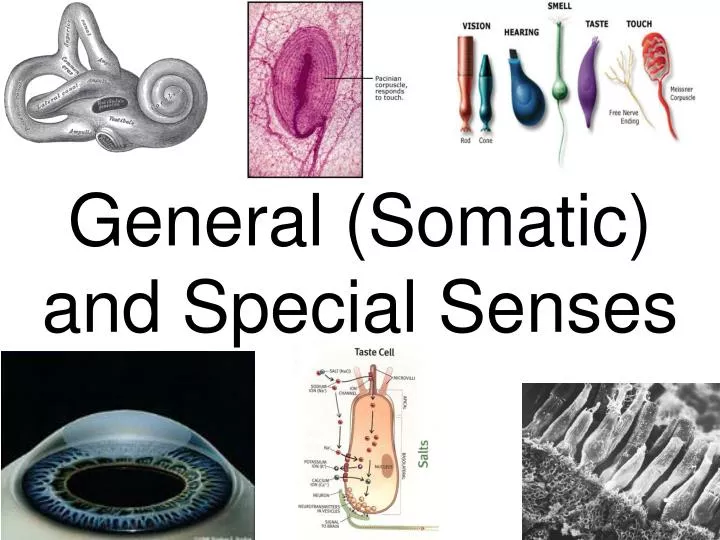
General (Somatic) and Special Senses. I. Receptors and Sensations. Sensory Receptors Detect change, trigger nerve impulses . Five general types Chemoreceptors Pain receptors Thermoreceptors Mechanoreceptors Photoreceptors. I. Receptors and Sensations. Sensations

E N D
I. Receptors and Sensations • Sensory Receptors • Detect change, trigger nerve impulses. • Five general types • Chemoreceptors • Pain receptors • Thermoreceptors • Mechanoreceptors • Photoreceptors
I. Receptors and Sensations • Sensations • Feelings that occur when the brain interprets sensory impulses. • Projection • Cerebral cortex sends the sensation back to its point of origin • Person can pinpoint the area of stimulation. • Sensory Adaptation • Impulses are sent at decreasing rates • Finally receptors fail to send impulses.
II. Sense of Sight • Visual Accessory Organs • Eyelid • Protects the eye • The thinnest skin of the body. • Lined with conjunctiva (folds back to cover eyeball). • Lacrimal Apparatus • Produces tears that lubricate and cleanse the eye. • Inferior and superior canaliculi drain tears into the nasal cavity (by nasalacrimal duct). • Tears also contain an antibacterial enzyme. • Extrinsic muscles of the eye attach to the sclera and move the eye.
II. Sense of Sight • Structure of the Eye (three distinct layers, or tunics) • The Outer Tunic (fibrous tunic) • Cornea • Transparent due to few cells and no blood vessels. • Helps focus light rays. • Sclera (white of eye) • Continuous with cornea. • Protects eye and is attachment for muscles. • The optic nerve and blood vessels pierce the sclera at the posterior of the eye.
II. Sense of Sight • The Middle Tunic (vascular tunic) • Choroid coat • Loosely joined to the sclera. • Highly vascular to nourish other tissues of the eye. • Darkly pigmented to keep the inside of the eye dark. • Ciliary body • Forms a ring around the front of the eye. • Composed of folds called ciliary processes and ciliary muscles.
II. Sense of Sight • Suspensory ligaments hold the lens in position and change its shape (focus). • Lens • Composed of epithelial cells called lens fibers. • The ability of the lens to adjust shape to facilitate focusing is called accommodation. • Iris • Colored portion of eye. • Adjusts the amount of light entering the pupil. • Has a circular set and a radial set of smooth muscle fibers.
II. Sense of Sight • Anterior cavity (two chambers) • Anterior chamber (between the cornea and iris) • Posterior chamber (between the iris and suspensory ligaments) • Filled with aqueous humor (from ciliary body). • Aqueous humor circulates from one chamber to the other through the pupil. • Too much aqueous humor causes glaucoma.
II. Sense of Sight • The Inner Tunic (Retina) • Covers the back side of the eye • Surrounds the posterior cavity • Filled with vitreous humor. • Composed of pigmented epithelium, visual receptor cells, and a layer of neurons
II. Sense of Sight • Macula lutea • Center of retina • Depression in middle is fovea centralis (the point of sharpest vision) • Optic disk • Medial to the fovea centralis • Your blind spot • Where nerve fibers leave the eye.
II. Sense of Sight • Light Refraction • Light waves must bend to be focused. • The cornea and lens bend light waves to focus them on the retina. • Myopia: nearsighted • Hyperopia: farsighted
II. Sense of Sight • Visual Receptors • Rods (elongated) • Function in dim light • Produce colorless vision. • Cones (blunt-shaped) • Provide sharp images in bright light • Enable us to see in color. • Highest concentration on the fovea centralis
II. Sense of Sight • Visual Pigments • Rhodopsin (in rods and cones) • Breaks down into a protein (opsin) and retinal (from vitamin A) in the presence of light. • Decomposition activates a nerve impulse. • Night blindness is caused by vitamin A deficiency.
II. Sense of Sight • Isodopsins (in cones) • Three types of cones. • Each sensitive to different wavelengths of light (red, green, blue) • All three sets stimulated, the color is white • None are stimulated, the color is black.
II. Sense of Sight • Visual Nerve Pathways • The axons of ganglion cells leave the eyes to form the optic nerves. • Fibers from the medial(nasal) half of the retina cross over in the optic chiasma. • Impulses are transmitted to the thalamus and then to the visual cortex of the occipital lobe.
III. Sense of Hearing • The ear provides the senses of hearing and equilibrium. • Human Range: 20-2000 Hz • External Ear • Auricle (pinna): collects sound waves • External auditory meatus (canal).
III. Sense of Hearing • Middle Ear • Begins with the tympanic membrane (eardrum) • Air-filled space (tympanic cavity) housing the 3 auditory ossicles. • Ossicles are the malleus, incus, and stapes. • Tympanic membrane vibrates the malleus, which vibrates the incus, then the stapes.
III. Sense of Hearing • The stapes vibrates the fluid inside the oval window of the inner ear. • Auditory ossicles both transmit and amplify sound waves. • Auditory Tube (eustachiantube) • Connects the middle ear to the throat. • Helps maintain equal air pressure on both sides of the eardrum.
III. Sense of Hearing • Inner Ear • An osseous labyrinth (canal) in the bone of the temporal bone. • A Membranous labyrinth is inside the osseous labyrinth. • Between the two labyrinths is perilymph (fluid). • Endolymph is inside the membranous labyrinth.
III. Sense of Hearing • The cochlea houses the organ of hearing • The semicircular canals function in equilibrium. • The oval window leads to the upper compartment, called the scala vestibuli. • The lower compartment is the scala tympani. • The cochlear duct lies between these two compartments • Duct is separated from the scala vestibuli by the vestibular membrane, and from the scala tympani by the basilar membrane.
III. Sense of Hearing • The Organ of Corti • Houses receptors called hair cells • Lies on the basilar membrane. • Hairs of cells extend into the endolymph of the cochlear duct. • Above the hair cells lies the tectorial membrane. • Sound waves make hairs rub against tectorial membrane stimulating receptor cells.
III. Sense of Hearing • Auditory Nerve Pathways • Epithelial receptor cells depolarize, allowing calcium to flood in. • Calcium forces vesicles to release neurotransmitters from cell base (no axons or dendrites). • Neurotransmitters stimulate sensory nerve fibers. • Nerve fibers carry impulses to the auditory cortices of the temporal lobes.
IV. Sense of Equilibrium • Consists of two parts: static and dynamic equilibrium. • Static Equilibrium • Determines the orientation of the head and body • Organs are the utricle and saccule (expansions of the membranous labyrinth). • A macula, consisting of hair cells and supporting cells, lies inside the utricle and saccule.
IV. Sense of Equilibrium • The hair cells contact gelatinous material holding otoliths (calcium carbonate stones). • Gravity causes the otoliths and gelatinous material to shift, bending hair cells and generating a nervous impulse. • Brain interprets as the position of the head.
IV. Sense of Equilibrium • Dynamic Equilibrium • Maintains balance when the head and body suddenly move and rotate. • Three semicircular canals detect rotational motion of the head • Ampulla are located in each semicircular canal • The organs of dynamic equilibrium are called cristae ampullaris and are located in the ampulla
IV. Sense of Equilibrium • Hair cells extend into a dome-shaped gelatinous cupula. • Rapid turning of the head or body generates impulses as the cupula bends hair cells
V. Sense of Smell • Olfactory Receptors • Olfactory receptors are chemoreceptors. • The senses of smell and taste operate together. • Olfactory Organs • Yellowish-brown masses in the upper nasal cavity. • Contain the olfactory receptors plus epithelial supporting cells.
V. Sense of Smell • Chemicals are first dissolved in the watery fluid of the nasal cavity. • Olfactory receptors are stimulated by chemicals. • Neurons carry the signal to the olfactory lobes.
V. Sense of Smell • Olfactory Stimulation • Each odor stimulates a set of specific protein receptors in cell membranes. • The brain interprets different receptor combinations as an olfactory code. • Olfactory receptors adapt quickly. • Anosmia is partial or complete loss of smell.
VI. Sense of Taste • Taste buds • Located within papillae of the tongue • Organs of taste • Scattered throughout the mouth and pharynx.
VI. Sense of Taste • Taste Receptors • Taste cells are modified epithelial cells that function as receptors. • Taste cells contain the taste hairs that are the portions sensitive to taste. • Chemicals must be dissolved in water (saliva) in order to be tasted. • Taste involves specific membrane protein receptors that bind with specific chemicals in food. • Taste receptors rapidly undergo adaptation.
VI. Sense of Taste • There are four types of taste cells. • Sweet receptors are plentiful near the tip of the tongue. • Sour receptors occur along the lateral edges of the tongue. • Salt receptors are abundant in the tip and upper portion of the tongue. • Bitter receptors are at the back of the tongue.
VII. General (Somatic) Senses • Receptors associated with the skin, muscles, joints, and viscera • Types • Touch • Pressure • Temperature • Pain
VIII. Touch and Pressure Senses • Sensory Nerve Fibers • In the epithelial tissues • Detect changes in pressure and touch • Meissner's Corpuscles • Flattened connective tissue sheaths. • Abundant in hairless areas • Sensitive to light touch • Pacinian Corpuscles • Large structures of connective tissue and cells • Detect deep pressure
VIII. Touch and Pressure Senses • Proprioceptors • Monitor body joint positions • In tendons and muscle • Baroreceptors- respond to blood pressure changes
IX. Temperature Senses • Heat Receptors and Cold Receptors (free nerve endings) • Both adapt quickly. • Temperatures near 45o C stimulate pain receptors • Temperatures below 10o C also stimulate pain receptors
X. Sense of Pain • Pain receptors • Free nerve endings that are stimulated when tissues are damaged • Adapt little, if at all. • None in nervous tissue of brain. • Visceral pain receptors are the only receptors in the viscera that produce sensations. • Referred Pain • Feels like it comes from elsewhere. • Due to common nerve pathways.
X. Sense of Pain • Pain Nerve Fibers • Conduct pain impulses away from their source. • Acute pain fibers • Thin, myelinated fibers. • Carry impulses rapidly and cease when the stimulus stops. • Chronic pain fibers • Thin, unmyelinated fibers. • Conduct impulses slowly and continue sending impulses after the stimulus stops.
X. Sense of Pain • Regulation of Pain Impulses • Aware of pain when impulses reach the thalamus. • Cerebral cortex mediates a response. • Brain can release presynaptic biochemicals which inhibit the pain impulses in the spinal cord. • Endorphins- In the pituitary and hypothalamus and provide natural pain control. • Serotonin- Stimulates other neurons to release enkalphins. • Enkalphins- Suppress acute and chronic pain (same receptors as morphine).