Download
1 / 5
50 likes | 215 Views
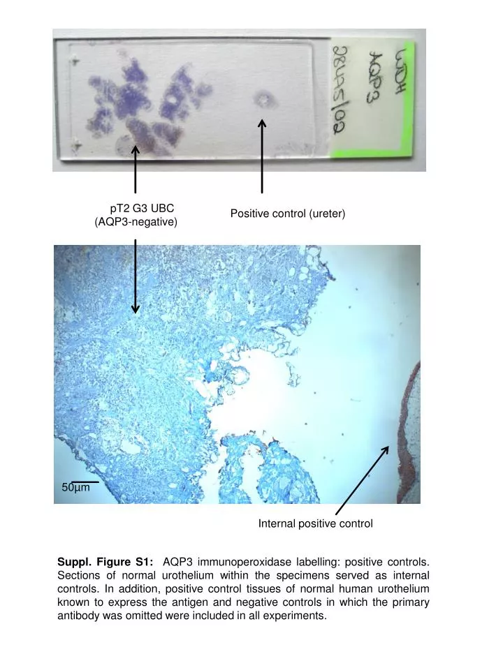
pT2 G3 UBC (AQP3-negative). Positive control ( ureter ). 50µm. Internal positive control.

E N D
pT2 G3 UBC (AQP3-negative) Positive control(ureter) 50µm Internal positive control Suppl. Figure S1: AQP3 immunoperoxidaselabelling: positive controls. Sections of normal urothelium within the specimens served as internal controls. In addition, positive control tissues of normal human urothelium known to express the antigen and negative controls in which the primary antibody was omitted were included in all experiments.
25µm Papillarylow-grade tumour (AQP3 +ve) Section of normal urothelium (internal positive control) Suppl. Figure S2: AQP3 immunoperoxidaselabelling. Intense, homogeneous AQP3 expression in >75% of a papillarylow-grade (pTaG1) tumour. Adjacent normal urotheliumshowingexpression in basal and intermediate, but not in superficialcells. No immunoreactivity was detected in the lamina propria, smooth muscle, or endothelium
AQP4, +vecontrol AQP4, pT1G2 AQP4 25µm AQP4, +vecontrol AQP4, pT2G3 Suppl. Figure S3: AQP4 immunoperoxidaselabelling. AQP4 was foundtobeexpressed in normal human urothelium but not in UC. Positive controls (human ureter) knownto express AQP4 wereincluded in all experiments.
-vecontrol (ureter) pT1 G2 25µm +vecontrole (ureter) pT2 G3 Suppl. Figure S4: AQP7 immunoperoxidaselabelling. AQP7 was expressed by all tumour specimens irrespective of grade and stage. Whereas AQP7 localized to the nucleus and the cytoplasm in normal human urothelium and superficial low-grade UC, intense labeling of the nuclei was found in muscle-invasive high-grade UC. Negative controls in which the primary antibody was omitted as well as positive controls known to express the antigen were included in all experiments.
+vecontrol (human liver) human ureter 25µm pTa G1 -vecontrol (human liver) Suppl. Figure S5: AQP9 immunoperoxidaselabelling. AQP 9 was showntobeintenselyexpressedby human liver (positive control), but noimmunoreactivity was detected in ureterand UC specimens.