Download

1 / 34
380 likes | 481 Views
Learn about the peritoneum, its layers, functions, and relations with different organs. Discover the roles of omentum, mesenteries, and ligaments in the abdominal cavity.

E N D
The peritoneum SHANDONG UNIVERSITY Liu Zhiyu
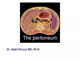
The peritoneum General features • The peritoneum is a thin serous membrane that line the walls of the abdominal and pelvic cavities and cover the organs within these cavities • Parietal peritoneum-lines the walls of the abdominal and pelvic cavities • Visceral peritoneum-covers the organs
The peritoneum • Peritoneal cavity -the potential space between the parietal and visceral layer of peritoneum. • In the mail, this is a closed cavity, but in female, there is communication with the exterior through the uterine tubes, the uterus, and the vagina
The peritoneum • Function • Secretes a lubricating serous fluid that continuously moistens the associated organs • Absorb • Support viscera
Relationship between viscera and peritoneum Intraperitoneal viscera • Viscera are almost totally covered with visceral peritoneum • Example • Stomach • Superior part of duodenum • Jejunum • Ileum • Cecum • Vermiform appendix • Transverse and sigmoid colons, • Spleen and ovary
Relationship between viscera and peritoneum Interperitoneal viscera • Most part of viscera surrounded by peritoneum • Example • Liver • Gallbladder • Ascending and descending colon • Upper part of rectum • Urinary bladder and uterus
Relationship between viscera and peritoneum Retroperitoneal viscera • Some organs lie on the posterior abdominal wall and are covered by peritoneum on their anterior surfaces only • Example • Kidney • Suprarenal gland • Pancreas • Descending and horizontal parts of duodenum • Middle and lower parts of rectum • Ureter
Structures which are formed by peritoneum • Omentum • Mesenteries • Ligaments • Folds, recesses or pouches
Omenta • Two-layered fold of peritoneum that connect the stomach with another viscus • Lessor omentum • Greater omentum
Lessor omentum • Two-layered fold of peritoneum which extends from porta hepatis to lesser curvature of stomach and superior part of duodenum • Hepatogastric ligament -extends from porta hepatis to lesser curvature of stomach • Hepatoduodenal ligament -extends from porta hepatis to superior part of duodenum. Contains: • Common bile duct • Proper hepatic a. • Hepatic portal v.
Lessor omentum Omental foramen • Behind the right border of hepatoduodenal ligament • Superior-caudate lobe of liver • Inferior-superior part of duodenum • Anteriorly-free border of hepatodudenal ligament • Posteriorly-peritoneum covering the inferior vena cava
Greater omentum • Four-layered fold of peritoneum • Connects the greater curvature of the stomach and superior part of duodenum with the transverse colon. • It hangs down like an apron in front of coils of small intestine and is folded back on itself. • If an infection occurs in the intestine, plasma cells formed in the lymph nodes combat the infection and help prevent it from spreading to the peritoneum.
Greater omentum Lesser omentum Lessor omentum
Omental bursa Position-lies behind the lesser omentum and stomach Walls • Superiorly-peritoneum which covers the caudate lobe of liver and diaphragm • Anteriorly-formed by lesser omentum, peritoneum of posterior wall of stomach, and anterior two layers of greater omentum • Inferiorly-conjunctive area of anterior and posterior two layers of greater omentum • Posteriorly-formed by posterior two layers of greater omentum, transverse colon and transverse mesocolon, peritoneum covering pancreas, left kidney and suprarenal gland
Omental bursa • Left • Spleen • gastrosplenic ligament • splenorenal ligament • Right-omental foramen • Communication The Omental bursa (lesser sac) communicates with the greater sac through the omental foramen.
Mesenteries • Two-layered fold of peritoneum connecting part of the intestines with the posterior abdominal wall
Mesenteries Mesentery • Two-layered fold of peritoneum suspends the small intestine from the posterior abdominal wall • Broad and a fan-shaped • Radix of mesentery • 15 cm long • Directed obliquely from left side of L2 to in front of right sacroiliac joint • Intestinal border-folded, 7 m long
Mesenteries Mesoappendix • Triangular mesentery-extends from terminal part of ileum to appendix • Appendicular artery runs in free margin of the mesoappendix
Mesenteries • Transverse mesocolon -a double fold of peritoneum which connects the transverse colon to the posterior abdominal wall • Sigmoid mesocolon -inverted V-shaped, with apex located in front of left ureter and division of common iliac artery
Ligaments • Two-layered folds of peritoneum that connect the lesser mobile solid viscera with the abdominal walls
Ligaments of liver • Falciform ligament of liver • Consists of double peritoneal layer • Extends from anterior abdominal wall (umbilicus) to live • Free border of ligament site of ligamentum teres
Ligaments of liver • Coronary ligament • Bare area of live • Left and right triangular ligaments • Hepatogastric ligament • Hepatoduodenal ligament • Ligamentum teres hepatis
Ligaments of spleen • Gastrosplenic ligament -a double layer of peritoneum that connects the fundus of stomach to hilum of spleen. In this double layer of peritoneum are the short gastric and left gastroepiploic vessels • Splenorenal ligament -extends between the hilum of spleen and anterior aspect of left kidney. The splenic vessels lies within this ligament, as well as the tail of pancreas • Phrenicosplenic ligament • Splenocolic ligament
Ligaments of stomach • Hepatogastric ligament • Gastrosplenic ligament • Gastrophrenic ligament • Gastrocolic ligament • Gastropancrestic ligament
Folds and recesses of posterior abdominal wall • Superior duodenal fold and recess • Inferior duodenal fold and recess • Intersigmoid recess -formed by the inverted V attachment of sigmoid mesocolon • Retrocecal recess -in which the appendix frequently lies
Folds and recesses of posterior abdominal wall • Hepatorenal recess -lies between the right lobe of liver, right kidney, and right colic flexure, and is the lowest parts of the peritoneal cavity when the subject is supine
Folds and fossas of anterior abdominal wall • Median umbilical fold -contain the remnant of urachus (median umbilical ligaments) • Medial umbilical fold-contains remnants of the umbilical arteries (medial umbilical ligaments) • Lateral umbilical fold -contains the inferior epigastric vessels • Supravesical fossa • Medial inguinal fossa • Lateral inguinal fossa
Pouches • In male-rectovesical pouch In female • Rectouterine pouch -between rectum and uterus • Vesicouterine pouch -between bladder and uterus
Peritoneal cavity subdivisions The transverse colon and transverse mesocolon divides the greater sac into: • Supracolic compartment (subphrenic space )-lies between diaphragm and transverse colon and transverse mesocolon • Infracolic compartment -lies between transverse colon and transverse mesocolon and pelvic inlet
Supracolic compartments Suprahepatic space • Right suprahepatic space • Right anterior suprahepatic space • Right posterior suprahepatic space • Bare area of live • Left suprahepatic space • Left anterior suprahepatic space • Left posterior suprahepatic space
Supracolic compartments Infrahepatic space • Right infrahepatic spacees (hepatorenal recess 肝肾隐窝) • Left infrahepatic space • left anterior infrahepatic space • left posterior infrahepatic space
Infracolic compartments • Right paracolic sulcus (gutter) • Lies lateral to the ascending colon. It communicates with the hepatorenal recess and the pelvic cavity • It provides a route for the spread of infection between the pelvic and the upper abdominal region • Left paracolic sulcus (gutter) • Lies lateral to the descending colon. • It is separated from the area around the spleen by the left phrenicocolic ligament, a fold of peritoneum that passes from the colic flexure to the diaphragm.
Infracolic compartments • Right mesenteric sinus -triangular space, lies between root of mesentery, ascending colon, right 2/3 of transverse colon and transverse mesocolon • Left mesenteric sinus -lies between root of mesentery, descending colon, right 1/3 of transverse colon and transverse mesocolon, its widens below where it is continuous with the cavity of the pelvis