Download
1 / 31
390 likes | 2.02k Views
Vascular Surgery. Angie Allen, ACNP Stacey Becker, RN . Objectives. Identify our team. Peripheral Artery Disease Cerebral Revascularization Lower Extremity Revascularization Lower Extremity Amputation Abdominal Aortic Aneurysms (endovascular) Thoracic Aortic Aneurysms (endovascular)

E N D
Vascular Surgery Angie Allen, ACNP Stacey Becker, RN
Objectives • Identify our team. • Peripheral Artery Disease • Cerebral Revascularization • Lower Extremity Revascularization • Lower Extremity Amputation • Abdominal Aortic Aneurysms (endovascular) • Thoracic Aortic Aneurysms (endovascular) • Abdominal Aortic Aneurysms (open) • Thoracic Outlet
Who are we? Attendings • Dr. Thomas Naslund-Division Chief • Dr. Raul Guzman
Who are We?Attendings Continued • Dr. Jeff Dattilo • Dr. Colleen Brophy
Who are we? • Fellows • Dr. Ali Khoobehi • Dr. Syed Rizvi • Interns: Carry the consult/resident pager: 831-6374
Who are we?Nurse Practitioner • Angie Allen, ACNP-BC First Call for Vascular M-F 0730-1600 886-0163 (cell) 835-8202 (pager)
Who are we? • Case Management Stacey Becker, RN (Dr. Naslund) Ann Luther, RN • Social Worker Ann Lacy, RN
Other Numbers • Vascular Office: 322-2343 • Vascular Clinic: 936-7485 • Vascular Lab: 343-9561
Arterial Disease • Peripheral Artery Disease (PAD): leading case of death worldwide. Polyvascular disease. • Atherosclerosis: Most likely the cause of PAD. Hardening of the artery or loss of elasticity. • Arterial Pathophysiology: 1. Occlusive disease: Atherosclerosis is symptomatic by gradually occluding the artery to the target organ or extremity. (kidneys, colon, legs, or arms) 2. Symptoms occur with critical arterial stenosis (75 % of cross sectional of lumen is obliterated)
Arterial Disease • Aneurysmal Disease: occurs due to loss of structural integrity of vessel wall. Over time this will result in dilation and aneurysm formation.
Cerebral Revascularization • Symptomatic: Patients who have carotid stenosis or occlusion that have exhibited a CVA or TIA • Asymptomatic: Patients who have carotid stenosis or occlusion that are high risk for CVA (i.e. hypertension, hyperlipidemia, smoker, obesity, CAD, etc.)
Symptoms • Right sided symptoms: -Left hemiplegia or monoparesis and right eye visual loss • Left sided symptoms: -Right hemiplegia or monoparesis and left eye visual loss -aphasia
Symptoms • Visual symptoms are due to ischemia of the retina. • Amaurosis fugax -Transient visual loss -”Window shade”, “flashing lights”, or “sparks”
Cerebral RevascularizationSurgical Intervention Carotid Endarterectomy Or Carotid Artery Stenting
Cerebral RevascularizationPost Operative Care • Neuro Assessment: VERY IMPORTANT. Essential for recognizing neurological deficits. • Contralateral hemiparesis: technical problem with endarterectomy with immediate return to OR. Notify team ASAP. Arterial duplex may be ordered. • Defuse neurological deficit: possible internal capsule stroke secondary to hypotensive episode. • Delayed neurological deficit: 12-24 hours postoperatively. Arterial Duplex with possible CTA of head and neck for evaluation of brain hemorrhage or CVA and evaluation of carotid.
Post Operative Care Continued • Dextran 40: instituted for antiplatelet purposes and may be continued for 24 hours postoperatively. • NPO until POD 1 for possible exploration. • D5 ½ NS while patient is NPO • POD 1: Initiation of Plavix 75 mg subcutaneous daily (if no concerns for hematoma) • Incision: Leave dressing dry and intact until POD 1, may remove. Incision will be closed with disolvable sutures, leave open to air unless draining.
Cerebral RevascularizationComplications • Hypertension: 20 % of patients. SBP 100-140 • Neck Hematoma: May compromise breathing and swallowing. -May require immediate surgical intervention for evacuation -Order tracheostomy kit Stat to the bedside • Local Nerve Injuries: Most common laryngeal and hypoglossal nerves presenting as temporary weakness in speech, swallowing, tongue or lip movement. Less than 0.5% result in permanent damage. • Hyperperfusion Syndrome: 1-2 % occur 3-7 days post operatively. Headache, Seizures, and Intracranial Hemorrhage. Hypertension may accompany. Supportive management
Cerebral VascularizationDischarge Instructions • Incision Care: Leave open to air, unless draining. Wash with antibacterial soap and water and use white wash cloths. • Immediately call 911 with patient has headache with associated decreased level of consciousness or seizure activities. • Follow up in Vascular Clinic 4 weeks postoperatively. • Discharge Medications: Plavix and pain medication • Plavix injection education. • Activity: Do not resume normal work activities until follow up apt. No driving until that time, do not return to work. (?????)
Lower Extremity Vascular DiseaseSymptoms • Claudication: pain at rest, present with ambulation. Typically seen one level below the disease. • Critical Ischemia: Rest pain may be first symptoms of severe ischemia. Sharp, localized pain to forefoot to below the ankle, dependent rubor and pallor with elevation. 95% loose limb in 1 yr without revascularization. • Critical Ischemia: Non healing ulcers. (arterial vs venous) • Critical Ischemia-Gangrene: Skin and subcutaneous tissue involvement. Dry (noninfected black eschar) vs Wet (macerated, purulent drainage).
Symptoms Continued • Microemboli: Blue Toe Syndrome causes blue, mottled spots over the toes. May be painful. • Acute Arterial Ischemia: Sudden onset of extremity pain, pallor, paresthesia, pulselessness, and poikilothermia. Caused by stenotic artery or emboli if no previous vascular disease.
TREATMENT • Treatment is based on duration, disability, progression, general medical condition, non-invasive diagnostic testing AND pathology • Non-op management: walking program, lifestyle modification, with possible medication. • Diagnostic Testing: Arterial duplex with segmental pressures/ABI’s (vascular lab), CTA or MRA, arteriogram, plain films, ECG (if ischemic toes-could be from a-fib), PT /PTT/INR/Platelet workup.
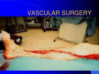
Operative Managment • Percutaneous transluminal angioplasty/stenting • Femoropopliteal or Pop-DP, etc. bypass (saphenous vein, Dakron, ePTFE) • Femoropopliteal percutaneous endovascular intervention • Aortoiliac or Aortobifemoral bypass or angioplasty with or without stenting • Thromboembolectomy • Amputation
Post-Operative Care • ICU stabilization after aortic operations (stability of vitals/hemodynamics, respiratory, fluid, electrolyte, cardiac, laboratory -pcv, blood glucose, lytes, coags- management). • Fluids: D51/2 NS 20 KCL at 75 mL/hr • Rewarm and vasodilate: bolus may be • warranted • Post op day 3-4: mobilization of fluids-may see lasix given.
Post-Operative Care Continued • Pain Control: essential for mobilization. PCA or percocet or lortab • Ambulation: PT/OT consult, POD 1 • Rooke Perioperative Boots • Antibiotics: continued for 24 hours • Wound Care: remove dressing POD 1, may leave open to air unless draining. Wash with antibacterial soap and water and use white wash cloths. • Amputation Wounds: Takedown is on POD 2, will require knee immobilizer. • High Risk for Pressure Ulcers
Complications • Hemorrhage from graft: Exploration required. • Thrombis (graft occlusion) PULSES< PULSES<PULSES • Infection Stage 1: Involving skin and dermis-wound care, antibiotics. Stage 2: Extending to subcutaneous and fatty tissue but not graft-Exploration and washout in the OR, continued wound care and antibiotics. Stage 3: Graft involvement-Exploration and washout in the OR with graft removal with establishment of new route of perfusion. Continued wound care and 6 weeks of IV antibiotics.
Complications Continued • Compartment Syndrome: Caused by prolonged ischemia (> 6 hrs) then revascularization resulting in edema in the calf muscles. Leg pain with sensory deficits to the dorsum of the foot and weakness of toe dorsiflexion. Measure Compartment Pressure. Treatment: fasciotomy.