Download
1 / 23
230 likes | 458 Views
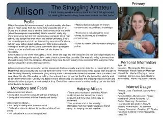
Allison Gong. Histology Microscopy Development. I. Histology. Histology. Histology = study of tissues Tissue = group of cells with similar structure and function Four types of human tissues: Nervous tissue Muscle Connective tissue Epithelial tissue. Connective tissue.

E N D
Allison Gong • Histology • Microscopy • Development
Histology • Histology = study of tissues • Tissue = group of cells with similar structure and function • Four types of human tissues: • Nervous tissue • Muscle • Connective tissue • Epithelial tissue
Connective tissue • Provides support (physical + metabolic) for other tissues • Surrounds all other tissues • Provides structural framework • Types of connective tissue: • Bone • Blood • Cartilage • Adipose tissue • Tendons, ligaments
Connective tissue • Combination of: • Cells (type varies) • Extracellular materials; matrix • Cells far apart • Matrix holds H2O • Resists compression • Nutrients/wastes pass through (interstitial fluid) • Stretchy and strong
Epithelial tissue • Covers surfaces, lines tubes, forms glands • Cells tightly packed, form sheets • Functions: • Protection (e.g., skin) • Absorption (e.g., gut) • Secretion (e.g., gut, exocrine/endocrine glands)
Epithelial tissue • Gland - group of epithelial cells, specialized to secrete specific substance(s) • Types of glands: • Exocrine gland - ducts; to outside of body • e.g., sweat gland • Endocrine gland - ductless; hormones distributed via bloodstream • e.g., thyroid, pancreas
Epithelial tissue Epithelial tissues are categorized by number and type (shape) of cells
Epithelial tissue • 3 cell types: • Cuboidal • Columnar • Squamous • Number of cells: • 1 layer thick - simple • Good for absorption (e.g., intestinal epithelium) • > 1 layer thick - stratified • Good for protection (e.g., skin)
Stratified cuboidal - sweat gland Epithelial tissue -stratified epithelia
Stratified columnar - duct Epithelial tissue -stratified epithelia
Stratified squamous - esophagus Epithelial tissue -stratified epithelia
Not usually visible with light microscope Functions: “glue” - holds tissues together Template for cell migration during development Separates epithelial and connective tissues: basement membrane connective tissue The basement membrane
Requirements for a clear image 1. Magnification - make the image larger than life-size 2. Resolution - ability to distinguish two objects in close proximity 3. Contrast - make the image stand out against background
1. Magnification • Our scopes have four objective lenses: • 4X • 10X • 40X • 100X (oil immersion only) • Ocular lenses are 10X • Total magnification = (objective)(ocular) e.g., if using the 40X objective, total magnification = (40)(10) = 400X
well resolved poorly resolved 2. Resolution i.e., How close can two objects be and still be seen as two objects? **Note: Increasing magnification does not help problems of resolution!!
3. Contrast • i.e., How well can you see the image against the background? • Living cells have little contrast • Use stains • Dyes that bind to certain functional groups in cells • Examples: hematoxylin, eosin • But, most stains kill cells • Use phase contrast lighting instead