Download

1 / 5
50 likes | 306 Views
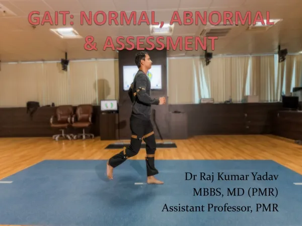
Anatomy of Normal Human Gait L. J. Rizzolo Yale University. Main Points. Need a language to discuss gait How we use a muscle differs from the anatomical description The language of the physical therapist facilitates discussion Fascial compartments organize our thinking

E N D
Anatomy of Normal Human Gait L. J. Rizzolo Yale University
Main Points • Need a language to discuss gait • How we use a muscle differs from the anatomical description • The language of the physical therapist facilitates discussion • Fascial compartments organize our thinking • A clinical approach focuses the discussion on a limited subset of muscles and nerves • A more comprehensive understanding can be built on this foundation Larry Rizzolo, Yale University
Types of Contraction Muscles can only pull by contraction --- They cannot push • Force of Deltoid > gravity Arm abductsconcentric contraction • Force of Deltoid = gravity Arm is stationary isocentric contraction • Force of Deltoid < gravity Arm adducts eccentric contraction Muscle Interactions Agonist vs Anatgonist Fine control of motion Synergist Modifies action by antagonizing unwanted motion Fixator Maintains position of one joint to enable movement of another joint Larry Rizzolo, Yale University
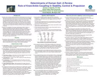
Femoral artery & nerve Obturator a. & n. Deep femoral a Sciatic Nerve Ant. Tibial a. Deep Peroneal n. Superficial Peroneal n. Post. Tibial a. Tibial n Peroneal a. Start simple: One compartment, one nerve Thigh • Which muscles to include? • Primary Clinicians focus on those that are: • Easy to test • Easy to see atrophy • Reporter for the rest of the compartment Leg Larry Rizzolo, Yale University
Exercises based on this approach http://info.med.yale.edu/surgery/anatomy Choose the <Exercises> link <lower limb> link Select from the exercises menu Larry Rizzolo, Yale University