Download
1 / 48
570 likes | 2.27k Views
CHAPTER 5 Function of Globular Proteins. Reversible binding of ligands Structure of myoglobin and hemoglobin Origin of cooperativity in hemoglobin Structure and function of antibodies Molecular mechanism of muscle movements. Key topics in protein function : . Functions of Globular Proteins.

E N D
CHAPTER 5Function of Globular Proteins • Reversible binding of ligands • Structure of myoglobin and hemoglobin • Origin of cooperativity in hemoglobin • Structure and function of antibodies • Molecular mechanism of muscle movements Key topics in protein function:
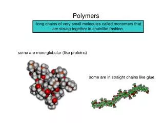
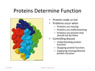
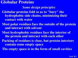
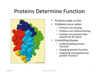
Functions of Globular Proteins • Storage of ions and molecules • myoglobin, ferritin • Transport of ions and molecules • hemoglobin, serotonin transporter • Defense against pathogens • antibodies, cytokines • Muscle contraction • actin, myosin • Biological catalysis • chymotrypsin, lysozyme
Interaction with Other Molecules • Reversible, transient process of chemical equilibrium: A + B AB • A molecule that binds is called a ligand (typically a small molecule) • A region in the protein where the ligand binds is called the binding site • Ligand binds via non-covalent forces, which enables the interactions to be transient
Binding: Quantitative Description • Consider a process in which a ligand (L) binds reversibly to a site in the protein (P) • The kinetics of such a process is described by: the association rate constant ka the dissociation rate constant kd • After some time, the process will reach the equilibrium where the association and dissociation rates are equal • The equilibrium composition is characterized by the the equilibrium constant Ka ka P L + L P kb
Spectroscopic Detection of Oxygen Binding to Heme • The heme group is a strong chromophore that absorbs both in UV and visible range • Ferrous form (Fe2+ ) without oxygen has an intense Soret band at 429 nm • Oxygen binding alters the electronic properties of the heme, and shifts the position of the Soret band to 414 nm • Binding of oxygen can be monitored by UV-Vis spectrophotometry • Deoxyhemoglobin (in venous blood) appears purplish in color and oxyhemoglobin (in arterial blood) is red http://omlc.ogi.edu/spectra/hemoglobin/
Binding: Analysis in Terms of the Bound Fraction • In practice, we can often determine the fraction of occupied binding sites • Substituting [PL] with Ka[L][P], we’ll eliminate [PL] • Eliminating [P] and rearranging gives the result in terms of equilibrium association constant: • In terms of the more commonly used equilibrium dissociation constant:
Binding: Graphical Analysis • The fraction of bound sites depends on the free ligand concentration and Kd • In a typical experiment, ligand concentration is the known independent variable • Kd can be determined graphically or via least-squares regression [L] [L]total
Binding: Thermodynamic Connections • Interaction strength can be expressed as: • association (binding) constant Ka, units M-1 • dissociation constant Kd, units M, Kd = 1/Ka • interaction (binding) free energy Go, units: kJ/mol Definitions: • Go = Ho -TSo:enthalpy and entropy • Ka = [PL]/[P][L] Kd=[P][L]/[PL] • Relationships: • Go = -RT ln Ka = RT ln Kd (RT at 25 oC is 2.48 kJ/mol) • Magnitudes • Strong binding: Kd < 10 nM • Weak binding: Kd > 10 M
Specificity: Lock-and-Key Model • Proteins typically have high specificity: only certain ligands bind • High specificity can be explained by the complementary of the binding site and the ligand. • Complementarity in • size, • shape, • charge, • or hydrophobic / hydrophilic character • “Lock and Key” model by Emil Fisher (1894) assumes that complementary surfaces are preformed. +
Specificity: Induced Fit • Conformational changes may occur upon ligand binding (Daniel Koshland in 1958). • This adaptation is called the inducedfit. • Induced fit allows for tighter binding of the ligand • Induced fit can increase the affinity of the protein for a second ligand • Both the ligand and the protein can change their conformations +
Function of Myoglobin • Need to store oxygen for metabolism • Protein side-chains lack affinity for O2 • Some transition metals bind O2 well but would generate free radicals if free in solution • Organometallic compounds such as heme are more suitable but Fe2+ in free heme could be oxidized to Fe3+ if two heme molecules share an oxygen molecule • Solution: • Capture the oxygen molecule with heme that is sequestered inside the protein • In mammals, myoglobin is the main oxygen storage protein
Spectroscopic Detection of Oxygen Binding to Heme • The heme group is a strong chromophore that absorbs both in UV and visible range • Ferrous form (Fe2+ ) without oxygen has an intense Soret band at 429 nm • Oxygen binding alters the electronic properties of the heme, and shifts the position of the Soret band to 414 nm • Binding of oxygen can be monitored by UV-Vis spectrophotometry • Deoxyhemoglobin (in venous blood) appears purplish in color and oxyhemoglobin (in arterial blood) is red http://omlc.ogi.edu/spectra/hemoglobin/
Binding of Carbon Monoxide • CO has similar size and shape to O2; it can fit to the same binding site • CO binds over 20,000 times better than O2 because the carbon in CO has a filled lone electron pair that can be donated to vacant d-orbitals on the Fe2+ • Protein pocket decreases affinity for CO, but is still binds about 250 times better than oxygen • CO is highly toxic as it competes with oxygen. It blocks the function of myoglobin, hemoglobin, and mitochondrial cytochromes that are involved in oxidative phosphorylation
Could Myoglobin Work as Good O2 Transporter? • pO2 in lungs is about 13 kPa: Mb binds oxygen well • pO2 in tissues is about 4 kPa: it will not release it! • Would lowering the affinity (P50) of myoglobin to oxygen help?
For Effective Transport Affinity Must Vary with pO2 • pO2 in lungs is about 13 kPa: it sure binds oxygen well • pO2 in tissues is about 4 kPa: it releases about half of it at pH 7.6
How Can Affinity to Oxygen Change Like This? • Must be a protein with multiple binding sites • Binding sites must be able to interact with each other • This phenomenon is called cooperativity • positive cooperativity: first binding event increases affinity at remaining sites • negative cooperativity: first binding event reduces affinity at remaining sites Positive cooperativity can be recognized by sigmoidal binding curves
Hill Plots – Hb and Mb Slope of the plot is proportional to n – the “order” of the ligand binding reaction
Hemoglobin • Hemoglobin is a tetramer of two subunits (a2b2) • Each subunit is similar to myoglobin
Subunit Interactions in Hemoglobin • PDB ID 1HGA
R and T States of Hemoglobin • T = Tense state, O2 binding triggers a T R conformational change • R = Relaxed state, this state has higher affinity for O2 than the T state • Deoxyhemoglobin subunits are more stable in the T state • Conformational change from the T state to the R state involves breaking ion pairs between the a1-b2 interface
Subunit Interactions: Details • Ion Pairs in the T state
pH and O2 binding to Hemoglobin • Blood in lungs has higher pH than blood in capillaries of metabolic tissues • Affinity for oxygen depends on the pH • Oxygen binds well at higher pH • Oxygen is released well at lower pH • The pH difference between lungs and metabolic tissues increases the O2 transfer efficiency • This is known as the Bohr effect
Hemoglobin and CO2 Export • CO2 is produced by metabolism in tissues and must be exported • Some CO2 exported as dissolved bicarbonate in the blood • Some CO2 is exported in the form of a carbamate on the amino terminal residues of each of the polypeptide subunits. • Notice that the formation of a carbamate yields a proton which can bind to hemoglobin and promote oxygen dissociation
Immunity • Innate (primitive, immediate, nonspecific) • Exists in most organisms, • triggered by • common bacteria • cellular damage signals • Adaptive immunity specific – vertebrate
Adaptive immunity • Cellular immune system • targets own cells that have been infected • also clears up virus particles and infecting bacteria • key players: Macrophages, killer T cells (Tc), and inflammatory T cells (TH1) • Humoral “fluid” immune system • targets extracellular pathogens • can also recognize foreign proteins • makes soluble antibodies • keeps “memory” of past infections • key players: B-lymphocytes and helper T-cells (TH2)
Cellular Immune System • Many infected cells display fragments of infectious particles on their surface • Phagocytes: specialized cells that eat invaders • Macrophages: large phagocytes that ingest bacteria
Humoral Immune System • Vertebrates also fight infections with soluble antibodies that specifically bind antigens • Antigens are substances that stimulate production of antibodies • Typically macromolecular in nature • Recognized as foreign by the immune system • Coat proteins of bacteria and viruses • Surface carbohydrates of cells or viruses • Antibodies are proteins that are produced by B cells and specifically bind to antigens • Binding will mark the antigen for destruction or interfere with its function • A given antibody will bind to a small region (epitope) of the antigen • One antigen can have several epitopes
Antibodies: Immunoglobulin G Fab: fragment, antigen binding Variable domain differs in antibodies from different B-cells
Muscle Structure • Muscle fiber: large, single, elongated, multinuclear cell • Each fiber contains about 1,000 myofibrils
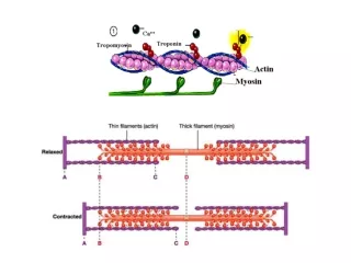
Myofibril Contains Thin and Thick Filaments • Thick filaments are aggregates of protein myosin • Thin filaments are aggregates of protein actin • Regulatory proteins modulate actino-myosin interactions
Myosin Thick Filaments Slide along Actin Thin Filaments • Cycle has four steps: • Myosin-ADP releases ADP, ATP binds; myosin dissociates from actin • ATP is hydrolyzed causing a conformational change of myosin into a high-energy state • Myosin in high-energy state re-connects to the actin filament • Power stroke where myosin returns to initial state; actin filament shifts relative to the myosin tail
Chapter 5: Summary In this chapter, we learned about: • how to quantitatively analyze binding data • how myoglobin stores oxygen • how hemoglobin transports O2, protons, and CO2 • how antibodies recognize foreign structures • how muscle works