Download
1 / 69
720 likes | 1.53k Views
. . . Indications for pulmonary artery catheterization in the ICU:Establish diagnosis of shock and/or respiratory failureGuide therapy of shock and/or respiratory failureBy improving oxygen delivery. . Oxygen delivery = CaO2 x COCardiac output = HR x SVSV is determined by:Preload (end-diastolic volume)Cardiac contractilityAfterload .

E N D
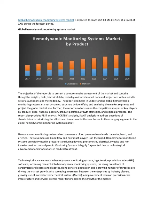
1. Hemodynamic monitoring All about the Swan
3. Indications for pulmonary artery catheterization in the ICU:
Establish diagnosis of shock and/or respiratory failure
Guide therapy of shock and/or respiratory failure
By improving oxygen delivery
4. Oxygen delivery = CaO2 x CO
Cardiac output = HR x SV
SV is determined by:
Preload (end-diastolic volume)
Cardiac contractility
Afterload
5. Information derived from PA catheter Directly measured
CVP
PAOP
Pulmonary artery pressure
SvO2
Cardiac output Calculated
Systemic vascular resistance
Pulmonary vascular resistance
Stroke volume
Oxygen delivery
6. Normal values Directly measured
CVP 2-4 mm Hg
PA 25/10
PAOP 8-12
SvO2 60-75%
Cardiac output 4-8 L/m
Cardiac index 2.5-4.0 L/min/M2
Calculated
SVR 900-1200 dynes sec/cm5
PVR 50-140
SV = 50-100mL
SV index 25-45
8. Insertion of Swan Ganz Ask why?
Then immediately ask why not:
Coagulopathy
Ventricular ectopy
LBBB
Pacemaker? Defibrillator?
Large pulmonary embolism
Severe pulmonary arterial hypertension Sustain VT (>30 beat run0 occurs in up to 3% pts
RBBB can occur in 5% catheter insertion
Pulmonary artery rupture: 30% mortality
Pulmonary artery pseudoaneurysmSustain VT (>30 beat run0 occurs in up to 3% pts
RBBB can occur in 5% catheter insertion
Pulmonary artery rupture: 30% mortality
Pulmonary artery pseudoaneurysm
9. Swan complications Associated with cordis placement
Ventricular arrhythmias requiring treatment 1.3 � 1.5%
Right bundle branch block ~0.5 -5%
Pulmonary artery rupture ~0.06 to 0.2%
Pulmonary artery pseudoaneurysm formation
Pulmonary infarction ~ 1.4%
Thromboembolic events ~1.6%
Mural thrombi
Sterile cardiac valve vegetation
Endocarditis esp of the pulmonic valve
10. So much information, why don�t we Swan more often? 1996 observational study
Swan within the first 24 hours of ICU admission associated with increased 30d hospital mortality (OR 1.24)
Association with poor outcome highest in the least sick pts
Meta-analysis of RCTs: no benefit but no harm
ESCAPE trial in patients with heart failure: no mortality benefit
RCT of peri-operative use in high risk pts undergoing cardiac, vascular or orthopedic surgery: no benefit
FACCT study of ARDS pts: no benefit of Swan v. CVP monitoring in managing vasoactive agents and fluid status
11. Nevertheless� PAC can be occasionally useful in the carefully selected patient
12. Insertion sites
13. Musts Full barrier precautions for maximal sterile technique
Flush and zero catheter prior to insertion at the phlebostatis axis
Remember catheter sheath
Once catheter tip is in the right atrium, always advance the catheter with the balloon inflated.
Always watch the waveforms transduced from the distal end of the catheter while advancing
Always withdraw catheter with the balloon deflated
Advance the catheter quickly while in the right ventricle
Advance slowly once the distal tip is in the pulmonary artery
18. Waveforms
24. Elevations in RAP Hypervolemia
Right ventricular infacrtion
Impaired RV contraction
Pulmonary hypertension
Pulmonic stenosis
Left to right shunts
Tricuspid valve disease
Cardiac tamponade
31. Overwedging
39. Abnormal waveforms
41. Seen with non compliant ventricle
Mitral or tricuspid stenosis
44. Seen with tricuspid valve regurgitation
Ventricular ischemia
Ventricular failure
Hypervolemia
47. Right ventricular pressure Peak systolic pressure
RV end-diastolic pressure
Early rapid filling (~60% of filling)
Slow phase (25% filling)
Atrial systolic phase
48. Left to right shunts Arterial sampling from RA, RV, and PA
Detection og an oxygen saturation �step-up� allows confirmation and determination of its location
Definition of �step-up� = >10% rise in oxygen saturation
49. Equalization of pressures RAP = RVed= PCWP
Cardiac tamponade
Constrictive pericardial disease
Restrictive cardiomyopathies
50. Cardiac output
51. Thermodilution Saline injected through the proximal port
Thermistor at the distal end of catheter measures the change in blood temperature over time
52. Area under the curve is inversely proportional to the rate of blood flow past the pulmonary artery
This rate is equivalent to cardiac output
53. Should not differ by more than 10%
54. Factors that decrease accuracy of thermodilution cardiac output Tricuspid regurgitation
Septal defects
Technical issues
Sensor malfunction
Improper injectate
55. Continuous thermodilution cardiac output 10 cm thermal filament located 15-25 cm from the catheter tip.
It generates low-energy head pulses transmitted to surrounding blood
56. Interpretation of the data
57. Cases
58. Case 1 20M presents post-MVA with abdominal pain.
T 97 BP 70/55 HR 130 RR 24
Exam: Alert, pale, diaphoretic. Extremities cool and clammy with poor capillary refill. Abdomen is distended and tender.
59. MAP = 60
CVP = 2
PA = 15/3
PAOP = 4
CO = 3
SvO2 50%
SVR?
SV?
What kind of shock?
60. Case 2 30F with flank pain, dysuria, fever to 104.
T 104 BP 70/35 HR 140
Exam: Flushed, warm, bounding pulses
61. MAP 47
CVP 2
PA 20/5
PAOP 5
CO 7
SvO2 75%
SV ?
SVR ?
What kind of shock?
62. Case 3 55M intermittent chest pains for last 24 hours presents with progressive shortness of breath and weakness
T 96 BP 80/60 HR 120 RR 28 SpO2 88%
Exam: Dyspneic, diaphoretic. Poor capillary refill. He has JVD, a gallop, soft murmur. Very little edema
63. MAP 67
CVP 10
PA 42/28
PAOP 29
CO 2.5
SvO2 55%
SV?
SVR?
What kind of shock?
64. Case 4 60M feeling bad and losing weight last 8 months. Hasn�t seen an MD in 30 years. Present with progressive weakness, shortness of breath, and edema.
T 96 BP 75/60 HR 120 RR 24 SpO2 92%
Exam: Cachectic. JVD. Distant heart sounds. Generalized edema. Thready pulses, poor capillary refill
65. MAP = 70
CVP 24
PA 40/24
PAOP 24
CO 2.4
SvO2 45%
SV?
SVR?
66. Case 5 46 F presents with worsening shortness of breath and chest pains over a 5 days period.
T 98 BP 78/62 HR 130 RR 28 pulse ox 84%
Exam: Tachypneic, dyspneic. JVD. Lungs clear. Heart sounds tacycardic with RV heave, pronounced S2, II/VI systolic murmur at LLSB.
67. MAP = 67
CVP 14
PA 60/28
PAOP 6
CO 3.5
SvO2 48%
SVR?
PVR?
SV? What is going on?
68. Case 6 36M admitted to the ICU with lobar pneumonia, septic shock.
Given 8 Liters of normal saline over 3 hours, but remains in refractory shock, requiring initiation of norephinephrine. Develops progressive hypoxemia and intubated. Post intubation CXR demonstrates bilateral pulmonary infiltrates
Exam T 103 BP 95/50 HR 120 RR 28 on vent SpO2 98%
Intubated, sedation. Warm and flushed with brisk capillary refill and bounding pulses.
69. MAP 65
CVP 9
PA 35/18
PAOP 16
CO 9.0
SvO2 80%
SVR?
SV?
Clinical scenario?