Download
1 / 70
710 likes | 1.19k Views
Learn about diagnostic modalities, complications, and management strategies for vascular injuries, including soft and hard signs, imaging studies, and surgical interventions, to ensure optimal patient outcomes.

E N D
VASCULAR INJURY Prof.Moaath AL Smady Jordan University Hospital
OUTLINE DIAGNOSTIC MODALITIES COMPLICATIONS COMPARTMENT ANATOMIC EXPOSURE FEMORAL Vs POPLITIAL Vs SHANK Vs
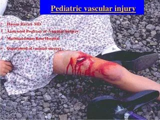
PEREPHIRAL VASCULAE INJURY • Distal to Deltopectoral Groove • Distal to Inginal ligament • Hard signs of Vascular injury needs Surgery
Hard Signs • Observed pulsatile bleeding • Ongoing heamorrhage with shock • Arterial thrill by manual palpation • Bruit over or near the artery • Abscent distal pulse • Signs of distal ischemia • Visible expanding hematoma
Soft Signs • Significant hemorrhage by History • Small non expanding heamatoma • Decreased pulse compared to the contralateral extremity • Bony injury • Wound proximity (1 cm from Vs) • Neurologic abnormality(anatomicaly related nerve)
Doppler ultrasound • Ankle Brachial Index • ABI < 0.90 = 87% sensitive, 97% specific for arterial injury • In absence of hard signs, can substitute this for screeningarteriography
Doppler ultrasound • Determine presence/absence of arterial supply • Assess adequacy of flow PRESENCE OF SIGNAL DOES NOT EXCLUDE ARTERIAL INJURY !
Imaging StudyDuplex US • Reliable for • Injury to arteries and veins • A-V fistulas • Pseudoaneurysms • Thrombosis • *It has 95% sensitivity &99 % specificity
Imaging study CT Angio * Ct angiograph faster, less expensive and less invasive 90-100 % sensitivity and 98% - 100 %specificity * diagnostic study of choice *Limitations: • difficulty differentiating spasm from occlusion • artifact from high attenuation structures like bullet fragments or other foreign matter
Management • ABCs • Active bleeding, limb threatening ischemia OR • Stable, good limb viability may investigate • Non-operative management non-occlusive lesion in asymptomatic patient • Pre-operative management • Prophylactic antibiotic • Single dose heparin iv if no C/I • Do not reperfuse dead limb! amputation
Immediate treatment • Control bleeding • Replace volume loss • Cover wounds • Reduce fractures/dislocations • Splint • Re-evaluate
Option of vascular repair • Arterial repair: (1) direct arterial repair (2) arterial patch repair (3) interposition graft repair (4) bypass repair (5) ligation • Venous repair whenever possible avoid ligation.
Tension-free primary repair Primary repair defect < 1-2 cm
Venous injury • Should be repaired in stable patient if technically feasible • Lateral venorrhaphy, EEA • Complex repair (PTFE, SVG) • Patency 75% • Before arterial repair • Limb threatening ischemia Shunt a. repair v. • Ligation is safe alternative esp. in unstable patients, complex injuries. • 86% free of edema at D/C Yelon, et al. J Trauma 1992 • DVT 78%, no significant sequelae of CVI Kurtoglu, et al. Am Surg 2007 • Long term anticoagulation? • early arterial graft failure
What is the management ? Mangled extremity :injury that involve al least ¾ consisting of bone ,soft tissue ,vessel,nerves
Amputation • Non-viable or non-savagable limb • Irreversible limb ischemia • Safe life before limbs!!! • Amputation can be life saving in life threatening extremity bleeding • Functional outcome consideration
Hemorrhage • Thrombosis • Infection • Stenosis • Miscellaneous
THROMBOSIS • most important complication • relatively common compared with other complications, • Perry et al found an early occlusion rate of 9.1%,
Inadequate arterial de´bridement • A second adjacent injury • Residual distal arterial thrombus • Severe stenosis at the suture line • Undue tension due to significant missing arterial segment • Twisting,or too long graft to cause a kink or external compression of the graft
INFECTION • Primary skin closure in a war wound • Placement of a vascular graft in an area of established infection • Inadequate soft tissue de´bridement in an attempt to conserve tissue for coverage of a vascular repair • Inadequate de´bridement of a damaged vessel
STENOSIS • Technical complication • Tight suture repair. • Lateral repair without sufficient remaining wall • Residual arterial wall damage. • Tension on the suture line
MISCELLANEOUS COMPLICATIONS Acute • Errors in diagnosis second associated or adjacent arterial injury Improper identification of the arteries may occur • Edema • Embolization • Disseminated intravascular coagulopathies
Delayed • Chronic pain Drapanas et al (1970)7 found that chronic pain was a complaint in10.2% • Decreased function • Ischemic changes • Systemic complications • Arteriovenous fistulas and false aneurysms • Arteriosclerotic changes • Aneurysmal graft changes
Compartment Syndrome • occurs when muscle swells within osteofacial compartment pressure exceed capillary pressure they end up with ischemia • Causes • Distal pulses • Distal nerve • Examination • Pressure
Pain, aggravated during stretching of the muscle group involved. • Pressure. • Paresthesia. • Paralysis, late manifestation • Pulselessness very late stages • Pallor
Measurement of Pressures and Laboratory Tests • Intracompartmental Pressure Monitoring 40 • Serum CreatinePhosphokinase and Myoglobin • This enzyme indicates muscle necrosis. • not appropriate for early detection, • useful to monitor the progression of equivocal syndromes or recently decompressed compartments. • Pulse Oximetry and Near-Infrared Spectroscopy
TREATMENT • Adequate skin incision and an adequate fascial incision • Pharmacologic Interventions • Mannitol
FasciotomyFasciotomy to fully decompress all involved compartments is the definitive treatment for ACS in the great majority of cases
INDICATIONS FOR FASCIOTOMY • Prolonged hypotension • Swelling of the extremity • Extensive soft tissue damage • Combined venous and arterial injury • Combined bony plus arterial or venous injury or both • Delay between injury and definitive repair • Compartmental pressure 35 mm Hg
70% of all arterial • More than 90% penetrating • most resulting from GSWs.12 • Injuries to the femoral artery are not commonly associated with fractures of the femoral shaft
OPERATIVE MANAGEMENT • proximal injuries it is wise to initially expose the distal common iliac vessels through a separate incision control before entering the femoral triangle. • The length of the sterile field includes the entire abdomen to the toes in both lower • bleeding can be controlled by direct pressure from the source of bleeding • Blind clamping is strongly discouraged