Download
1 / 41
410 likes | 522 Views
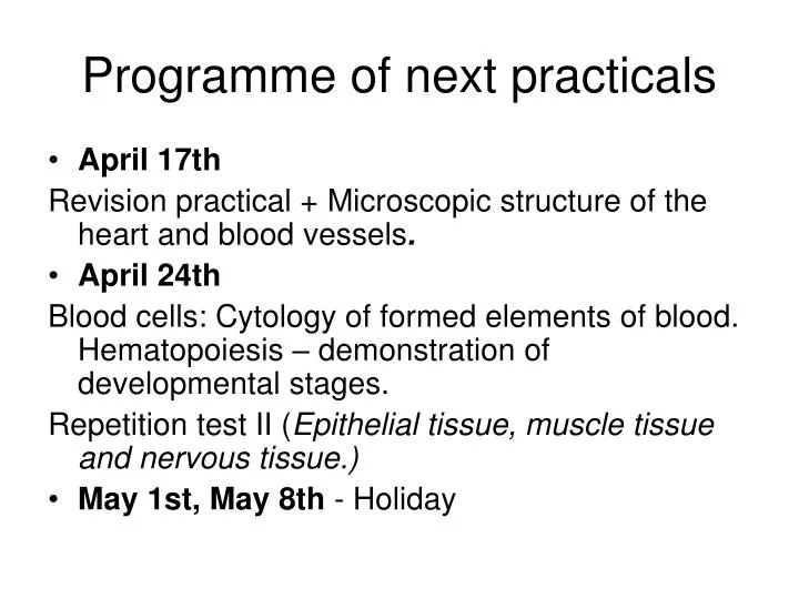
Programme of next practicals. April 17th Revision practical + Microscopic structure of the heart and blood vessels . April 24th Blood cells: Cytology of formed elements of blood. Hematopoiesis – demonstration of developmental stages.

E N D
Programme of next practicals • April 17th Revision practical + Microscopic structure of the heart and blood vessels. • April 24th Blood cells: Cytology of formed elements of blood. Hematopoiesis– demonstration of developmental stages. Repetition test II (Epithelial tissue, muscle tissue and nervous tissue.) • May 1st, May 8th - Holiday
General histology: Repetition Tissue types • Connective and supporting (+blood) • Epithelial • Muscular • Nervous
Connective tissue developed from mesenchyme consists of: • cells • intercellular matrix: • amorphous ground substance • fibers
Classification • connective tissue proper • specialized connective (supporting) tissue: cartilage bone Functions • mechanical (cartilage, bone) • nutritional (intercellular substance) • defensive (cells: histiocytes, plasma cells, leukocytes – immunocompetence, production of antibodies)
Connective tissue proper • mucous (jelly-like) • loose collagenous (areolar) • dense collagenous (regular, irregular) • reticular • elastic • adipose tissue (white, brown)
Connective tissue proper Mucous (jelly-like) connective tissue
Cartilage composed of cells (chondrocytes) and intercellular matrix: fibers and amorphous substance (chondrocytes present in lacunae within the matrix) fibers: • collagenous only • combination of collagenous and elastic cartilage is nonvascular, without nerves perichondrium – dense irregular connective tissue on the surface: important for growth and nutrition of cartilage Classification according to the kind and amount of fibers: • hyaline cartilage • elastic cartilage • fibrocartilage
Bone • Cells: osteocytes, osteoblasts, osteoclasts, osteoprogenitor cells • Intercellular matrix: collagenous fibers (type I), amorphous substance, inorganic salts • Macroscopically - 2 types: compact (dense) and spongy (cancelous) • Microscopically – 2 types according to the organisation of intercellular substance: woven (nonlamellar) and Haversian (lamellar)
Epithelial tissue Classification on the structural basis (arrangement of cells): • membranes – cells form sheets – the most common type, including most exocrine glands • trabecular – cells are arranged into anastomosing trabeculae – liver, endocrine glands • reticular – stellate cells form a network – thymus Classification on the basis of function : • Covering (lining) epithelia – epithelial membranes • Glandular epithelium • Absorptive epithelium – enterocytes (intestine) • Respiratory epithelium – pneumocytes (lung) • Sensory epithelium – olfactory ep., taste buds • Myoepithelial cells (exocrine glands, m. dilatator pupillae)
Covering epithelia (epithelial membranes) • Simple squamous
Covering epithelia (epithelial membranes) • Simple cuboidal
Covering epithelia (epithelial membranes) • Simple columnar
Covering epithelia (epithelial membranes) • pseudostratified
Covering epithelia (epithelial membranes) • Stratified squamous nonkeratinized
Covering epithelia (epithelial membranes) • Stratified squamous keratinized
Covering epithelia (epithelial membranes) • Stratified columnar
Covering epithelia (epithelial membranes) • transitional
Glandular epithelium Unicellular glands Goblet cells Paneth cells
Glandular epitheliummulticellular glands – ducts and secretory portions (acini, tubules)
Glandular epithelium • Multicellular glands – serous acini and mucous tubules
Muscle tissue Morphological unit of: • Skeletal muscle cell is called muscle fiber – rhabdomyocyte (multinucleated, nuclei at periphery) myofibrils are structures inside the cell, consist of myofilaments (actin, myosin) • Cardiac muscle cell – cardiomyocyte (uninucleated, nucleus centrally) myofibrils, intercallated discs • Smooth muscle cell – leiomyocyte (uninucleated, nucleus centrally) no myofibrils, only myofilaments
Nervous tissue Anatomically: • CNS (central nervous system): brain, spinal cord • PNS (peripheral nervous system): nerves, ganglia Histologically it consists of 2 principal cell types: • nerve cells (neurons) – excitability (irritability) and conductivity • supporting cells (neuroglia)