Download
1 / 33
561 likes | 1.5k Views
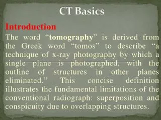
CT BASICS AND CT BRAIN. HISTORY. Sir Godfrey hounsfield-1972 Nobel prize in 1979 with cormack six generation of scanners Latest 128 multidetector ct. PRINCIPLE. Internal structure of an object can be reconstructed from multiple projections of the object.

E N D
HISTORY • Sir Godfrey hounsfield-1972 • Nobel prize in 1979 with cormack • six generation of scanners • Latest 128 multidetector ct
PRINCIPLE • Internal structure of an object can be reconstructed from multiple projections of the object. • Uses x rays applied in sequence of slices across the organ • Images reconstructed from xray absortion data • Xray beam moves around the patient in a circular path
PARTS 1)xray tube-akin to that in a x ray machine. 2)detectors 3)gantry- which houses xray apparatus 4)patient couch 5)viewing console
HOW A CT SCAN IS DONE • A motorized table moves the patient through the CT imaging system • a source of x-rays rotates within the circular opening, and a set of x-ray detectors rotates in synchrony on the far side of the patient. • In axial CT, which is commonly used for head scans, the table is stationary
In helical CT, which is commonly used for body scans, the table moves continuously as the x-ray source and detectors rotate, producing a spiral or helical scan • Data processed by computer to form image
TYPES • Spiral ct- • uses principle of volumetric acquisiton. • no respiratory misregistration • EBCT-coronary calcium measurement • HRCT • CT cisternography and myelography
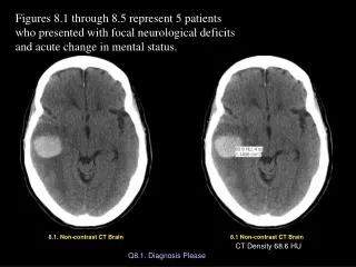
INDICATIONS • Acute changes in mental status • Focal neurologic findings • Trauma • Suspected SAH • Initial evaluation of conductive hearing loss
CT • Advantages – • Easy availabilty • Fast • Better for bone and acute blood,lesions of skull base and calvarium • Calcification • Less limited by patient factors • Disadvantages- • high radiation • poor visualisation of posterior fossa lesions
CT DENSITY MEASUREMENT • Hounsfield units • Water-0HU • Air- -1000 HU • Calcification- +1000HU • Fat-100HU • CSF-3HU • Grey matter-38HU • White matter-30HU • Fresh blood-70-80HU
CECT • To detect abnormal disrution caused by tumor,abscess ,infarct etc • Uses ionic or non ionic contrast(6 fold reduction in allergic reactioin 0.04%) • In normal CNS vessels,pituitary choroid and dura enhance
Indications for non ionic contrast • Prior adverse reaction • BA • Allergy or atopy hx • <2yr • RF(Cr>2) • Cardiac • DM • Severe debilitation
INTERPRETATION OF CT BRAIN • 1-GENERAL INFORMATION • 2-EXTRACRANIAL TISSUE • 3-CRANIAL BONE • 4-BLOOD • 5-CSF FLOW • A-VENTRICULAR SYSTEM • B-CISTERNS • 6-BRAIN TISSUE • A-MASS LESIONS • B-SULCI & GYRI • C-GRY & WHITE DIFFERENTIATION
I-EXTRACRANIAL TISSUE II-CRANIAL BONES
LV V3
Physiologic calcifications • Chorid plexus-rare before 10yrs • Basal ganglia-rare before 40ys • Pineal gland-common after 30 yr rare before 10yr • Falx • Dentate nuclei